. 2008 Dec 11;51(23):7405-16.
doi: 10.1021/jm800483v.

A small molecule inhibitor, 1,2,4,5-benzenetetraamine tetrahydrochloride, targeting the y397 site of focal adhesion kinase decreases tumor growth

Affiliations

A small molecule inhibitor, 1,2,4,5-benzenetetraamine tetrahydrochloride, targeting the y397 site of focal adhesion kinase decreases tumor growth

Vita M Golubovskaya et al. J Med Chem. .

Abstract

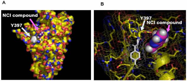
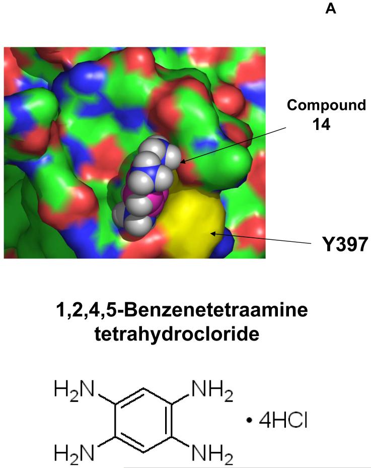
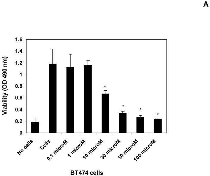
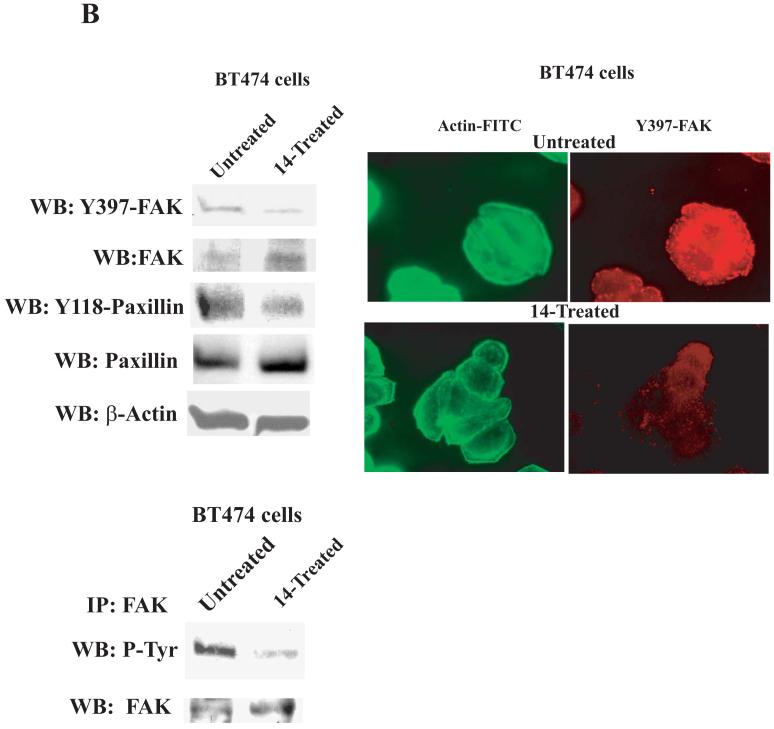
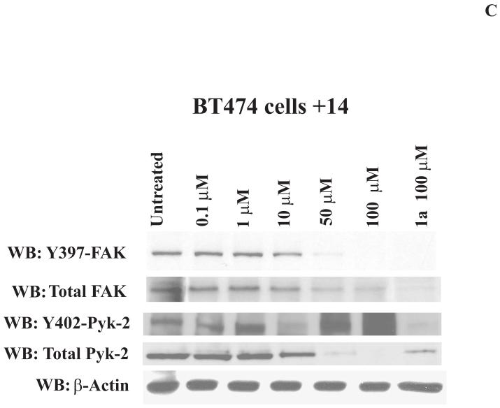
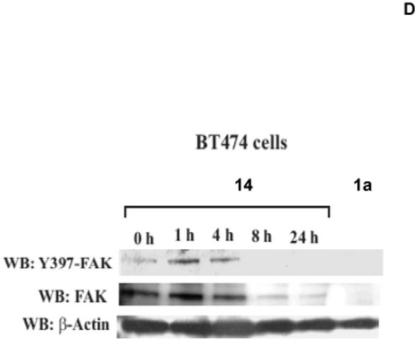
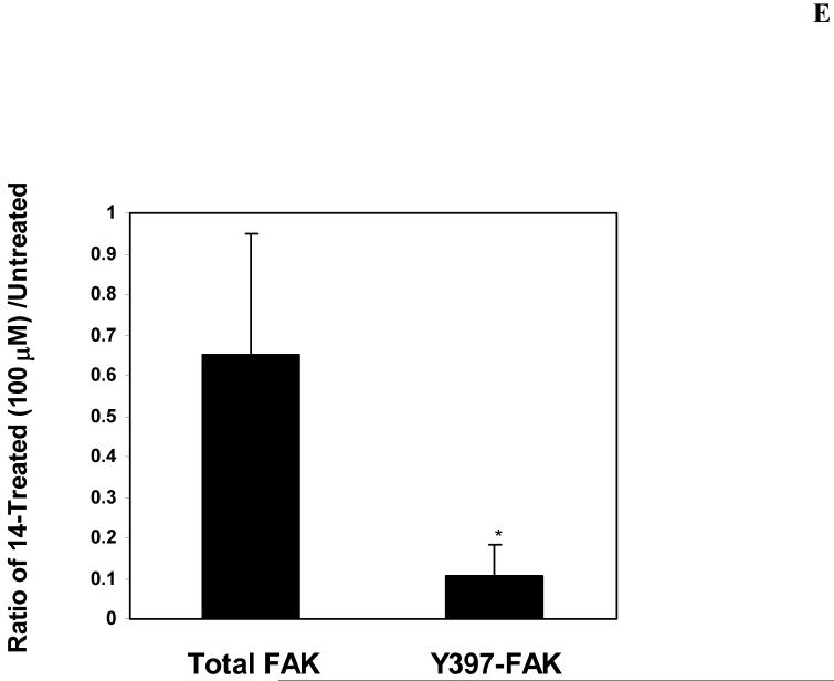
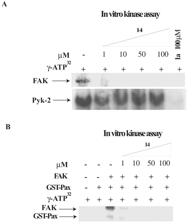
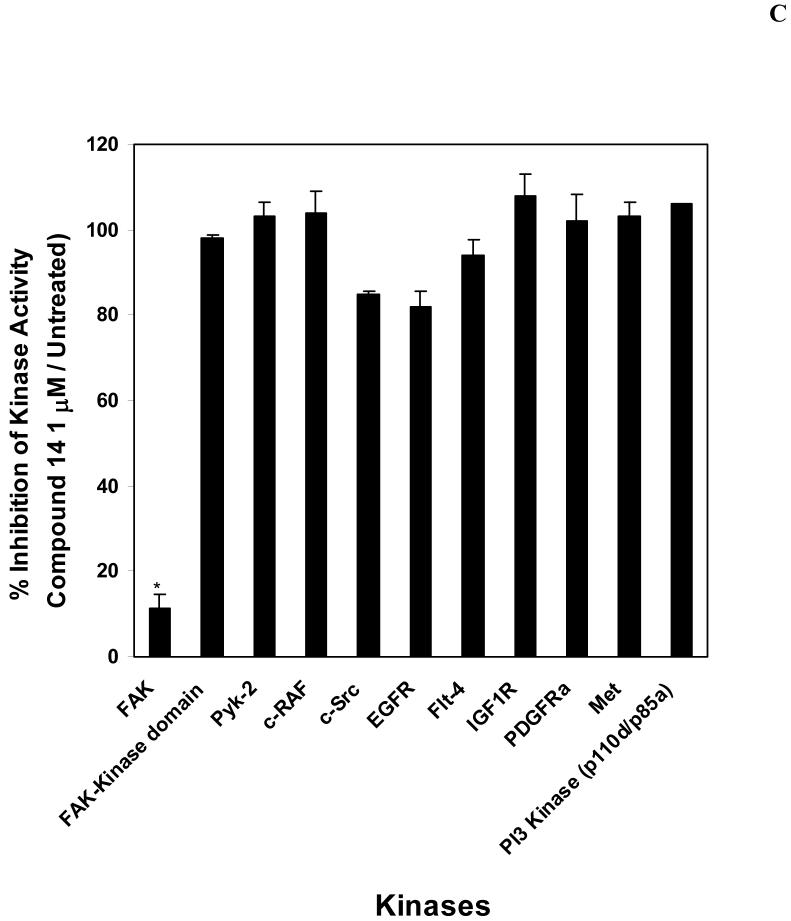
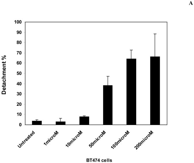
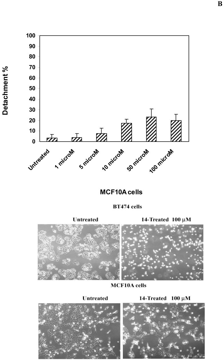
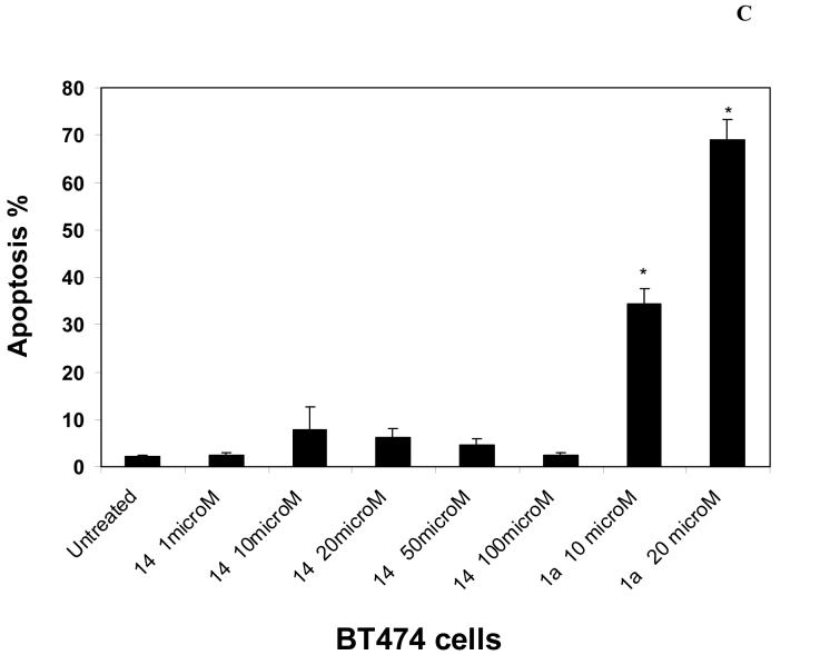
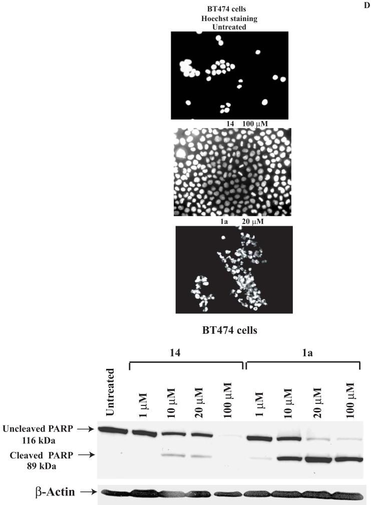
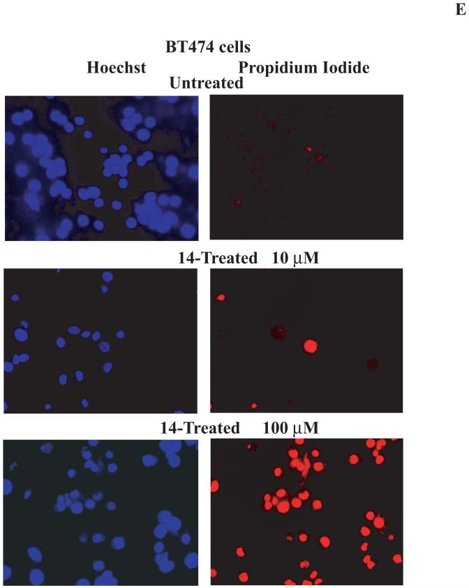
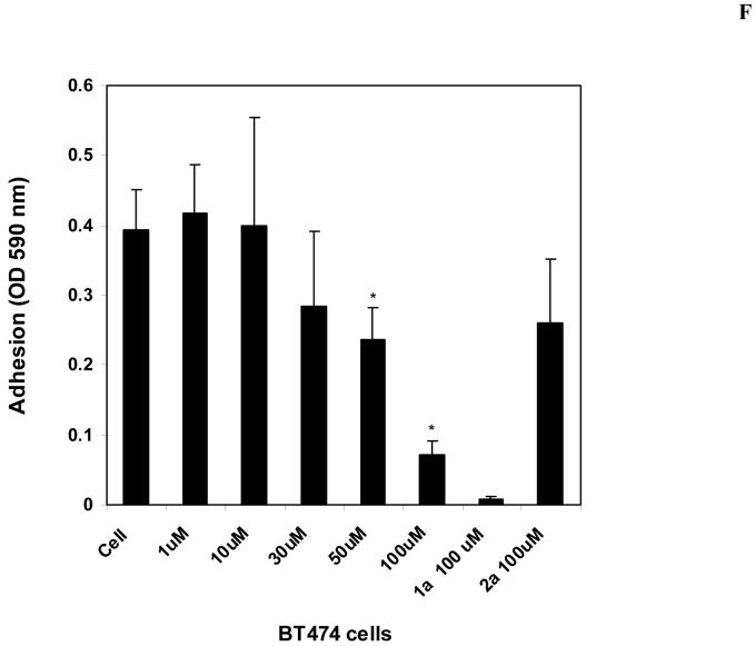
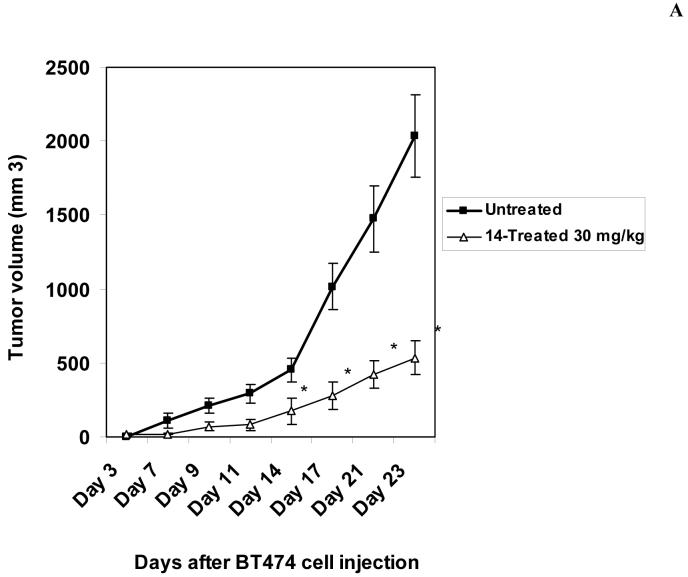
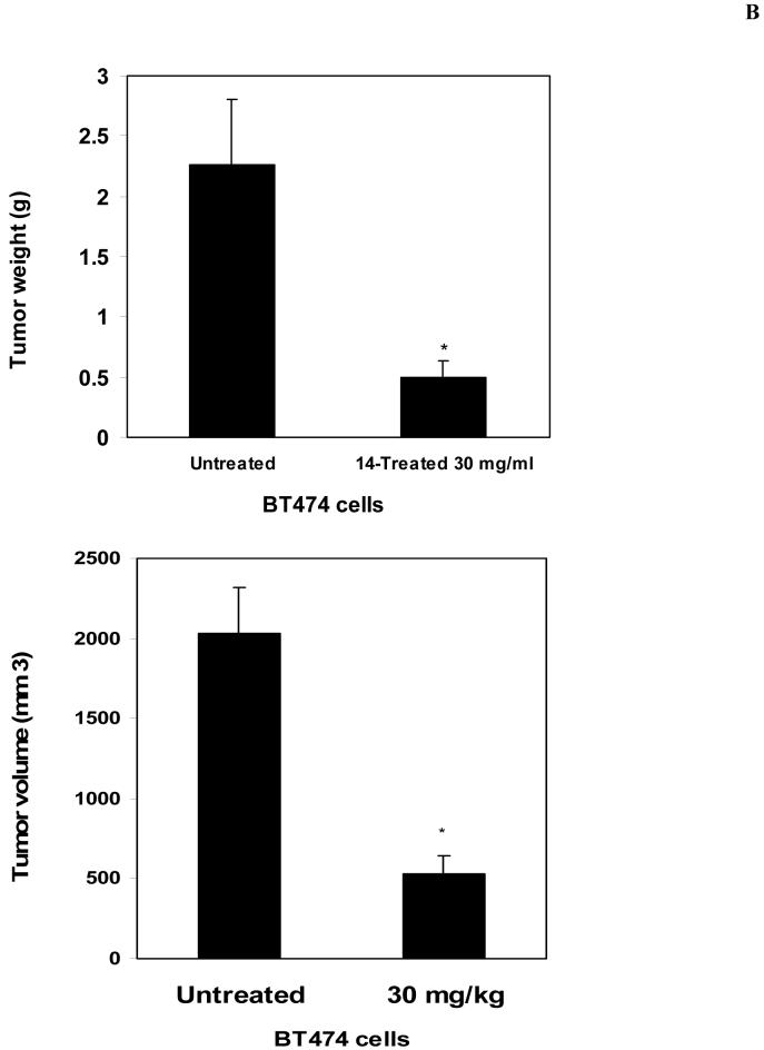
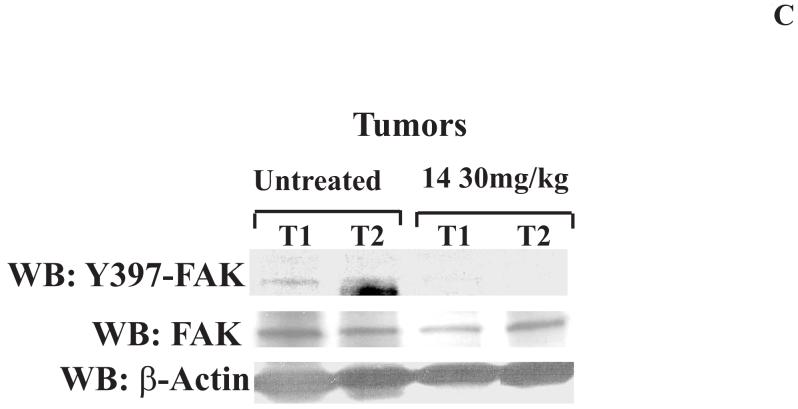
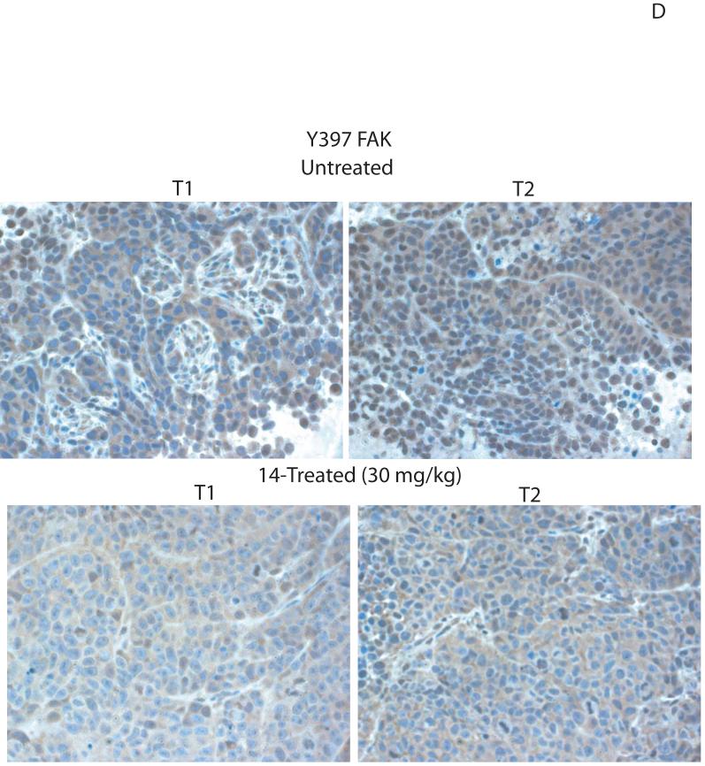
Focal adhesion kinase (FAK) is a nonreceptor kinase that is overexpressed in many types of tumors. We developed a novel cancer-therapy approach, targeting the main autophosphorylation site of FAK, Y397, by computer modeling and screening of the National Cancer Institute (NCI) small molecule compounds database. More than 140,000 small molecule compounds were docked into the N-terminal domain of the FAK crystal structure in 100 different orientations that identified 35 compounds. One compound, 14 (1,2,4,5-benzenetetraamine tetrahydrochloride), significantly decreased viability in most of the cells to the levels equal to or higher than control FAK inhibitor 1a (2-[5-chloro-2-[2-methoxy-4-(4-morpholinyl)phenylamino]pyrimidin-4-ylamino]-N-methylbenzamide, TAE226) from Novartis, Inc. Compound 14 specifically and directly blocked phosphorylation of Y397-FAK in a dose- and time-dependent manner. It increased cell detachment and inhibited cell adhesion in a dose-dependent manner. Furthermore, 14 effectively caused breast tumor regression in vivo. Thus, targeting the Y397 site of FAK with 14 inhibitor can be effectively used in cancer therapy.


Figures

Figure 1
Figure 1
A, B. Targeting of Y397 site of FAK by structure-based molecular docking approach. A, The crystal structure of FAK (FERM) domain, reported in with one of the NCI compound targeting Y397 site of FAK (shown by arrows). B, Zoomed image of Y397 site and this example compound (B). C, D. The effect of compounds targeting Y397 site on viability of breast cancer and melanoma cell lines. C, The 35 compounds, 1-35 (Table 1) were added to the cells for 24 hours at 100 μM dose and MTT assay was performed, as described in Materials and Methods. The known FAK inhibitor, 1a (Novartis), was used as a control. T47D breast cancer cell line (C) and C8161 (D) melanoma cell line. Bars show means ± standard deviations. Compound 14 decreased maximally cancer cell viability (shown by arrow).
Figure 1
Figure 1
A, B. Targeting of Y397 site of FAK by structure-based molecular docking approach. A, The crystal structure of FAK (FERM) domain, reported in with one of the NCI compound targeting Y397 site of FAK (shown by arrows). B, Zoomed image of Y397 site and this example compound (B). C, D. The effect of compounds targeting Y397 site on viability of breast cancer and melanoma cell lines. C, The 35 compounds, 1-35 (Table 1) were added to the cells for 24 hours at 100 μM dose and MTT assay was performed, as described in Materials and Methods. The known FAK inhibitor, 1a (Novartis), was used as a control. T47D breast cancer cell line (C) and C8161 (D) melanoma cell line. Bars show means ± standard deviations. Compound 14 decreased maximally cancer cell viability (shown by arrow).
Figure 2
Figure 2
A. The structure of 14 compound. Upper panel: Compound 14 targets Y397 site of FAK. Lower panel: Chemical structure and name of the 14 compound. B. The closeup view of compound 14 interaction in the Y397 site of FAK. The secondary structure of alpha helices and beta sheets of the FAK-N-terminal domain are shown. 14 compound is shown in purple. The red dashed lines show hydrogen bonds and blue dashed line shows hydrophobic interaction. Y397 residue is shown in orange.
Figure 2
Figure 2
A. The structure of 14 compound. Upper panel: Compound 14 targets Y397 site of FAK. Lower panel: Chemical structure and name of the 14 compound. B. The closeup view of compound 14 interaction in the Y397 site of FAK. The secondary structure of alpha helices and beta sheets of the FAK-N-terminal domain are shown. 14 compound is shown in purple. The red dashed lines show hydrogen bonds and blue dashed line shows hydrophobic interaction. Y397 residue is shown in orange.
Figure 3
Figure 3
A, B, C. The compound 14 inhibits cell viability and decreases Y397 FAK phosphorylation in a dose-dependent manner. A, BT474 breast cancer cells were treated with different doses of 14 inhibitor for 24 hours and MTT assay was performed to test the effect on cell viability. Bars show mean of triplicate determinations ± standard deviations. 14 inhibits cell viability in a dose-dependent manner. *P<0.05 viability of 14-treated cells versus control untreated cells. B, Left upper panel: 14 specifically inhibits Y397 phosphorylation of FAK. BT474 cells were treated with 14 at 100 μM for 24 hours and Western blotting with Y397 and Y118 paxillin antibodies was performed to detect the level of phosphorylated FAK and paxillin, respectively. Western blotting with total FAK, paxillin and β-Actin was performed to detect expression of proteins in the cells. 14 effectively inhibited phosphorylation of Y397 and FAK substrate, Y118-paxillin. B, Right upper panel: Immunostaining with Y397-FAK antibody was performed on BT474 cells either untreated or treated with 14 at 100 μM for 24 hours. Immunostaining was performed with primary Y397-FAK antibody and Rhodamine-conjugated secondary antibody. FITC-BodipyFL-phallacidin was used for actin staining. 14 significantly decreased Y397-FAK in 14-treated cells. B, left lower panel: 14 blocked total phosphorylation of FAK. Immunoprecipitation was performed with FAK antibody and Western blotting with phosphotyrosine antibody was performed. The blot was stripped and probed with FAK antibody. 14 blocked phosphorylation of FAK. C, 14 decreases Y397 phosphorylation in a dose-dependent manner. Cells were treated with different doses of 14 inhibitor and Western blotting was performed with Y-397 and then with FAK antibody. Western blotting with Y402-Pyk-2 antibody was performed to detect Y402-Pyk-2 phosphorylation. Western blotting with β-Actin antibody was performed to control equal protein loading. D. The compound 14 inhibits FAK autophosphorylation in a time-dependent manner. Cells were treated with 100 μM of 14 inhibitor for 1, 1, 4, 8 and 24 hours. Treatment with 1a inhibitor at 100 μM for 24 hours was used as a control. Western blotting with Y397 was performed to detect Y397-FAK level. Then the blot was stripped and Western blotting with FAK and β-Actin was performed. The compound 14 inhibits Y397-FAK phosphorylation in a dose-dependent manner. E. Quantification of inhibition of Y397-phosphorylation, caused by 14 compound. Densitometry analysis was performed of 14-treated and untreated cells at 100 μM dose at three independent experiments using Scion Image, NIH software. Total FAK and Y397-FAK were expressed relatively to β-Actin, and then ratio of total FAK and Y397-FAK levels was calculated in -treated cells and expressed relative to untreated cells. Bars show mean ratio of 14-treated/untreated cells ± standard errors. * p<0.05 Y397-FAK compared to total FAK.
Figure 3
Figure 3
A, B, C. The compound 14 inhibits cell viability and decreases Y397 FAK phosphorylation in a dose-dependent manner. A, BT474 breast cancer cells were treated with different doses of 14 inhibitor for 24 hours and MTT assay was performed to test the effect on cell viability. Bars show mean of triplicate determinations ± standard deviations. 14 inhibits cell viability in a dose-dependent manner. *P<0.05 viability of 14-treated cells versus control untreated cells. B, Left upper panel: 14 specifically inhibits Y397 phosphorylation of FAK. BT474 cells were treated with 14 at 100 μM for 24 hours and Western blotting with Y397 and Y118 paxillin antibodies was performed to detect the level of phosphorylated FAK and paxillin, respectively. Western blotting with total FAK, paxillin and β-Actin was performed to detect expression of proteins in the cells. 14 effectively inhibited phosphorylation of Y397 and FAK substrate, Y118-paxillin. B, Right upper panel: Immunostaining with Y397-FAK antibody was performed on BT474 cells either untreated or treated with 14 at 100 μM for 24 hours. Immunostaining was performed with primary Y397-FAK antibody and Rhodamine-conjugated secondary antibody. FITC-BodipyFL-phallacidin was used for actin staining. 14 significantly decreased Y397-FAK in 14-treated cells. B, left lower panel: 14 blocked total phosphorylation of FAK. Immunoprecipitation was performed with FAK antibody and Western blotting with phosphotyrosine antibody was performed. The blot was stripped and probed with FAK antibody. 14 blocked phosphorylation of FAK. C, 14 decreases Y397 phosphorylation in a dose-dependent manner. Cells were treated with different doses of 14 inhibitor and Western blotting was performed with Y-397 and then with FAK antibody. Western blotting with Y402-Pyk-2 antibody was performed to detect Y402-Pyk-2 phosphorylation. Western blotting with β-Actin antibody was performed to control equal protein loading. D. The compound 14 inhibits FAK autophosphorylation in a time-dependent manner. Cells were treated with 100 μM of 14 inhibitor for 1, 1, 4, 8 and 24 hours. Treatment with 1a inhibitor at 100 μM for 24 hours was used as a control. Western blotting with Y397 was performed to detect Y397-FAK level. Then the blot was stripped and Western blotting with FAK and β-Actin was performed. The compound 14 inhibits Y397-FAK phosphorylation in a dose-dependent manner. E. Quantification of inhibition of Y397-phosphorylation, caused by 14 compound. Densitometry analysis was performed of 14-treated and untreated cells at 100 μM dose at three independent experiments using Scion Image, NIH software. Total FAK and Y397-FAK were expressed relatively to β-Actin, and then ratio of total FAK and Y397-FAK levels was calculated in -treated cells and expressed relative to untreated cells. Bars show mean ratio of 14-treated/untreated cells ± standard errors. * p<0.05 Y397-FAK compared to total FAK.
Figure 4
Figure 4
A, B. The compound 14 directly blocks in vitro catalytic autophosphorylation and kinase activity of FAK. A, upper panel. 14 blocks FAK catalytic autophosphorylation activity. In vitro kinase assay was performed with γ-ATP32, 0.1 μg of purified recombinant FAK protein and different doses of 14 inhibitor for 10 minutes at room temperature, as described in Materials and Methods. 14 directly blocks FAK autophosphorylation activity in a dose-dependent manner. A, lower panel. 14 has no inhibiting effect on Pyk-2 catalytic autophosphorylation activity. In vitro kinase assay was performed with γ-ATP32, 0.1 μg of purified recombinant Pyk-2 protein and different doses of 14 inhibitor for 10 minutes at room temperature, as described in Materials and Methods. 14 did not affect significantly Pyk-2 autophosphorylation. The data are from three independent experiments. B. The compound 14 blocks FAK kinase activity in vitro. GST-paxillin was added as a substrate in the kinase assay reaction. The reaction was performed with γ-ATP32, 0.1 μg of purified recombinant FAK protein and different doses of 14 inhibitor for 10 minutes at room temperature. 14 inhibits phosphorylation of paxillin in a dose-dependent manner. C. Kinase profile of compound 14. The compound 14 was screened against nine commercially available recombinant enzymes and catalytically active FAK kinase domain (411-686 a.a.) without the N-terminal domain, containing Y397 site by Kinase Profiller Service (Millipore/Upstate Biotechnology Inc.). The assay was performed with 1 μM dose of 14 that inhibited activity of FAK (Figure 4 A and B), 10 μM ATP and kinase substrates as described in Materials and Methods (http://www.millipore.com/drugdiscovery/dd3/KinaseProfiler). 14 effectively blocks full length FAK kinase activity (Figure 4A, B) and it doesn’t affect activities of homologous Pyk-2, FAK kinase domain and other kinases. The bars show percentage of inhibition of the enzyme activity (14/Untreated). The mean and standard errors of two independent experiments are shown.
Figure 4
Figure 4
A, B. The compound 14 directly blocks in vitro catalytic autophosphorylation and kinase activity of FAK. A, upper panel. 14 blocks FAK catalytic autophosphorylation activity. In vitro kinase assay was performed with γ-ATP32, 0.1 μg of purified recombinant FAK protein and different doses of 14 inhibitor for 10 minutes at room temperature, as described in Materials and Methods. 14 directly blocks FAK autophosphorylation activity in a dose-dependent manner. A, lower panel. 14 has no inhibiting effect on Pyk-2 catalytic autophosphorylation activity. In vitro kinase assay was performed with γ-ATP32, 0.1 μg of purified recombinant Pyk-2 protein and different doses of 14 inhibitor for 10 minutes at room temperature, as described in Materials and Methods. 14 did not affect significantly Pyk-2 autophosphorylation. The data are from three independent experiments. B. The compound 14 blocks FAK kinase activity in vitro. GST-paxillin was added as a substrate in the kinase assay reaction. The reaction was performed with γ-ATP32, 0.1 μg of purified recombinant FAK protein and different doses of 14 inhibitor for 10 minutes at room temperature. 14 inhibits phosphorylation of paxillin in a dose-dependent manner. C. Kinase profile of compound 14. The compound 14 was screened against nine commercially available recombinant enzymes and catalytically active FAK kinase domain (411-686 a.a.) without the N-terminal domain, containing Y397 site by Kinase Profiller Service (Millipore/Upstate Biotechnology Inc.). The assay was performed with 1 μM dose of 14 that inhibited activity of FAK (Figure 4 A and B), 10 μM ATP and kinase substrates as described in Materials and Methods (http://www.millipore.com/drugdiscovery/dd3/KinaseProfiler). 14 effectively blocks full length FAK kinase activity (Figure 4A, B) and it doesn’t affect activities of homologous Pyk-2, FAK kinase domain and other kinases. The bars show percentage of inhibition of the enzyme activity (14/Untreated). The mean and standard errors of two independent experiments are shown.
Figure 5
Figure 5
A. The compound 14 causes dose-dependent cell detachment in BT474 cells. BT474 cells were treated with different doses of 14 inhibitor. The detachment was determined on a hemacytometer, as described in Materials and Methods. Bars show means ± standard errors in three independent experiments. 14 significantly decreased cell detachment. Bars show means ± standard errors in three independent experiments. *P<0.05 versus untreated cells. B. Detachment caused by 14 in normal breast MCF10A cells. Upper panel: Normal breast MCF10A cells were treated with different doses of 14 inhibitor. The detachment was determined, as described in Figure 5A. Bars show means ± standard errors in three independent experiments. Lower panel: A phase-contrast microscopy of MCF10A cells treated with 14 at 100 μM dose. Detachment is shown in BT474 and MCF10A cells treated with 100 μM of 14. No significant detachment was caused by 14 in MCF10A cells compared with BT474 cells. C. The compound 14 doesn’t cause significant apoptosis in BT474 cells. Hoechst staining was performed on BT474 cells at 24 hours treatment with different doses of 14 and 1a inhibitors, as described in Materials and Methods. No significant apoptosis was detected with 14 compared to 1a inhibitor. Bars represent means ± standard deviations in four independent experiments. *P<0.05 versus untreated cells. D. Upper panel: Hoechst staining of 14-treated BT474 cells. BT474 cells treated as above were analyzed by Hoechst staining. Apoptotic nuclei stained with Hoechst are shown. No apoptotic nuclei were observed with 14 inhibitor at 100 μM dose compared to 1a inhibitor at 20 μM dose. Lower panel: PARP cleavage in 14-treated and 1a-treated cells. Western blotting on BT474 cells, treated with 14 or 1a inhibitors at different doses was performed with PARP antibody. 1a caused significantly higher levels of cleaved PARP (89 kDa) compared with 14-treated cells. No cleaved 89 kDa PARP was detected at 100 μM dose in 14-treated cells compared with 1a-treated cells. E. Propidium iodide staining of 14-treated cells. BT474 cells treated with 14 at 10 and 100 μM as above, were collected and stained for 10 minutes with 10 μg/ml of propidium iodide, detecting dead necrotic cells and with Hoechst (10 μg/ml), detecting nuclei. Cells were analyzed under the Zeiss fluorescent microscope. Propidium iodide staining was increased in a dose-dependent manner and reached maximum at 100 μM dose. F. The compound 14 blocks cell adhesion in a dose-dependent manner. BT474 cells were treated with 14 at different concentration and cell adhesion was measured as described in Materials and Methods. 1a and 2a inhibitors at 100 μM were used as a control. 14 significantly blocked cell adhesion in a dose dependent manner. Bars show means ± standard errors in four independent experiments*, P<0.05, cells adhesion in 14 treated cells less than in control untreated cells.
Figure 5
Figure 5
A. The compound 14 causes dose-dependent cell detachment in BT474 cells. BT474 cells were treated with different doses of 14 inhibitor. The detachment was determined on a hemacytometer, as described in Materials and Methods. Bars show means ± standard errors in three independent experiments. 14 significantly decreased cell detachment. Bars show means ± standard errors in three independent experiments. *P<0.05 versus untreated cells. B. Detachment caused by 14 in normal breast MCF10A cells. Upper panel: Normal breast MCF10A cells were treated with different doses of 14 inhibitor. The detachment was determined, as described in Figure 5A. Bars show means ± standard errors in three independent experiments. Lower panel: A phase-contrast microscopy of MCF10A cells treated with 14 at 100 μM dose. Detachment is shown in BT474 and MCF10A cells treated with 100 μM of 14. No significant detachment was caused by 14 in MCF10A cells compared with BT474 cells. C. The compound 14 doesn’t cause significant apoptosis in BT474 cells. Hoechst staining was performed on BT474 cells at 24 hours treatment with different doses of 14 and 1a inhibitors, as described in Materials and Methods. No significant apoptosis was detected with 14 compared to 1a inhibitor. Bars represent means ± standard deviations in four independent experiments. *P<0.05 versus untreated cells. D. Upper panel: Hoechst staining of 14-treated BT474 cells. BT474 cells treated as above were analyzed by Hoechst staining. Apoptotic nuclei stained with Hoechst are shown. No apoptotic nuclei were observed with 14 inhibitor at 100 μM dose compared to 1a inhibitor at 20 μM dose. Lower panel: PARP cleavage in 14-treated and 1a-treated cells. Western blotting on BT474 cells, treated with 14 or 1a inhibitors at different doses was performed with PARP antibody. 1a caused significantly higher levels of cleaved PARP (89 kDa) compared with 14-treated cells. No cleaved 89 kDa PARP was detected at 100 μM dose in 14-treated cells compared with 1a-treated cells. E. Propidium iodide staining of 14-treated cells. BT474 cells treated with 14 at 10 and 100 μM as above, were collected and stained for 10 minutes with 10 μg/ml of propidium iodide, detecting dead necrotic cells and with Hoechst (10 μg/ml), detecting nuclei. Cells were analyzed under the Zeiss fluorescent microscope. Propidium iodide staining was increased in a dose-dependent manner and reached maximum at 100 μM dose. F. The compound 14 blocks cell adhesion in a dose-dependent manner. BT474 cells were treated with 14 at different concentration and cell adhesion was measured as described in Materials and Methods. 1a and 2a inhibitors at 100 μM were used as a control. 14 significantly blocked cell adhesion in a dose dependent manner. Bars show means ± standard errors in four independent experiments*, P<0.05, cells adhesion in 14 treated cells less than in control untreated cells.
Figure 6
Figure 6
A, B. The compound 14 significantly blocks tumor growth in vivo. A, BT474 breast cancer cells were subcutaneously injected in 5 mice. The day after injection, 14 compound at 30 mg/kg was added daily 5 days per week. Five untreated mice were used as a control group. Tumor volume was measured with calipers. 14 significantly blocked tumor growth in vivo. Bars represent means ± standard deviations (n=5) P<0.05, Student’s t-test. B, The compound 14 significantly blocks tumor weight and volume. At day 23 after breast cancer cell injection, tumors were extracted, and weight and volume was determined in grams (upper panel) and in mm3 (lower panel), respectively. 14 significantly blocked tumor weight (upper panel) and volume (lower panel). Bars represent means ± standard deviations. * p<0.05, Student’s t-test. C, D. The compound 14 decreases Y397-FAK phosphorylation in tumors. C, We isolated tumors from untreated mice and from mice, treated with 14 compound. Cell lysates were prepared, and Western blotting was performed with Y397 antibody. FAK and β-actin antibodies were used for detecting total FAK and β-Actin levels. D, Immunohistochemical staining analysis was performed on untreated and 14-treated tumors with Y397-antibody. Two representative tumors from untreated and 14 treated mice groups are shown (marked as T1 and T2 tumors in each group) (C, D). 14 decreased Y397FAK phosphorylation in tumors treated with 14 (30 mg/kg) compared with untreated mice.
Figure 6
Figure 6
A, B. The compound 14 significantly blocks tumor growth in vivo. A, BT474 breast cancer cells were subcutaneously injected in 5 mice. The day after injection, 14 compound at 30 mg/kg was added daily 5 days per week. Five untreated mice were used as a control group. Tumor volume was measured with calipers. 14 significantly blocked tumor growth in vivo. Bars represent means ± standard deviations (n=5) P<0.05, Student’s t-test. B, The compound 14 significantly blocks tumor weight and volume. At day 23 after breast cancer cell injection, tumors were extracted, and weight and volume was determined in grams (upper panel) and in mm3 (lower panel), respectively. 14 significantly blocked tumor weight (upper panel) and volume (lower panel). Bars represent means ± standard deviations. * p<0.05, Student’s t-test. C, D. The compound 14 decreases Y397-FAK phosphorylation in tumors. C, We isolated tumors from untreated mice and from mice, treated with 14 compound. Cell lysates were prepared, and Western blotting was performed with Y397 antibody. FAK and β-actin antibodies were used for detecting total FAK and β-Actin levels. D, Immunohistochemical staining analysis was performed on untreated and 14-treated tumors with Y397-antibody. Two representative tumors from untreated and 14 treated mice groups are shown (marked as T1 and T2 tumors in each group) (C, D). 14 decreased Y397FAK phosphorylation in tumors treated with 14 (30 mg/kg) compared with untreated mice.

Similar articles

Cited by

References

    1. Schaller MD. The focal adhesion kinase. J Endocrinol. 1996;150(1):1–7. - PubMed
    1. Hildebrand JD, Schaller MD, Parsons JT. Identification of sequences required for the efficient localization of the focal adhesion kinase, pp125FAK, to cellular focal adhesions. Journal of Cell Biology. 1993;123(4):993–1005. - PMC - PubMed
    1. Xing Z, Chen HC, Nowlen JK, Taylor SJ, Shalloway D, Guan JL. Direct interaction of v-Src with the focal adhesion kinase mediated by the Src SH2 domain. Mol Biol Cell. 1994;5(4):413–21. - PMC - PubMed
    1. Cobb BS, Schaller MD, Leu TH, Parsons JT. Stable association of pp60src and pp59fyn with the focal adhesion-associated protein tyrosine kinase, pp125FAK. Molecular & Cellular Biology. 1994;14(1):147–55. - PMC - PubMed
    1. Schaller MD, Hildebrand JD, Shannon JD, Fox JW, Vines RR, Parsons JT. Autophosphorylation of the focal adhesion kinase, pp125FAK, directs SH2-dependent binding of pp60src. Molecular & Cellular Biology. 1994;14(3):1680–8. - PMC - PubMed

Publication types

MeSH terms

Substances