

Selectivity and mechanism of action of a growth factor receptor-bound protein 2 SRC homology 2 domain binding antagonist
- PMID: 18989951
- PMCID: PMC2651228
- DOI: 10.1021/jm800523u
Selectivity and mechanism of action of a growth factor receptor-bound protein 2 SRC homology 2 domain binding antagonist
Abstract
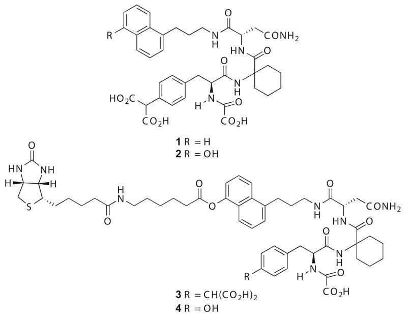
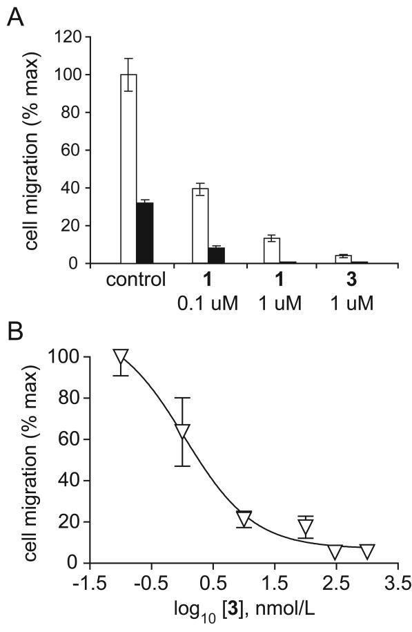
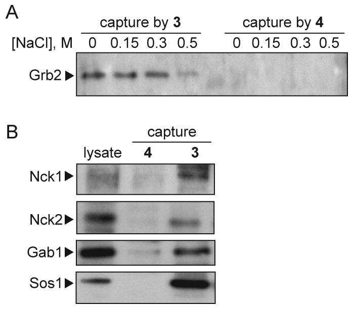
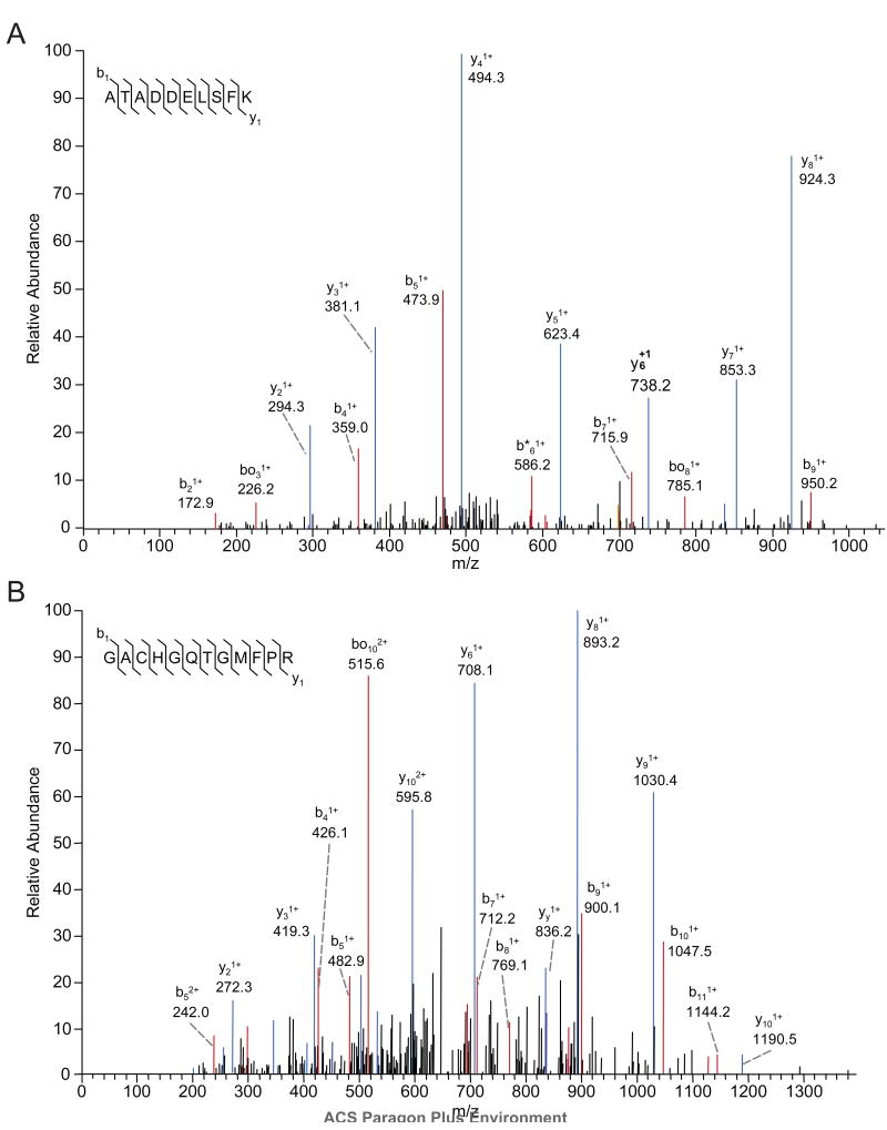
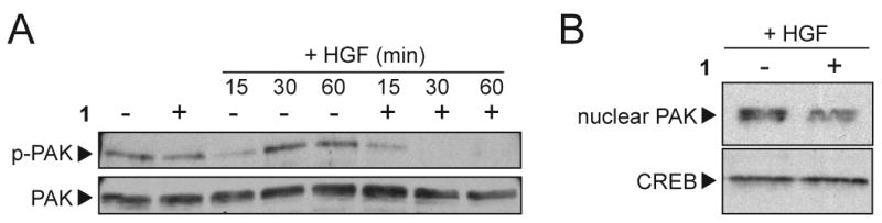
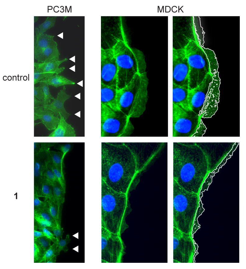
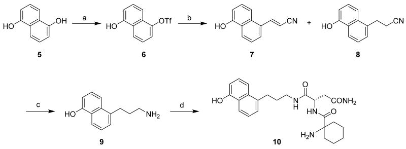
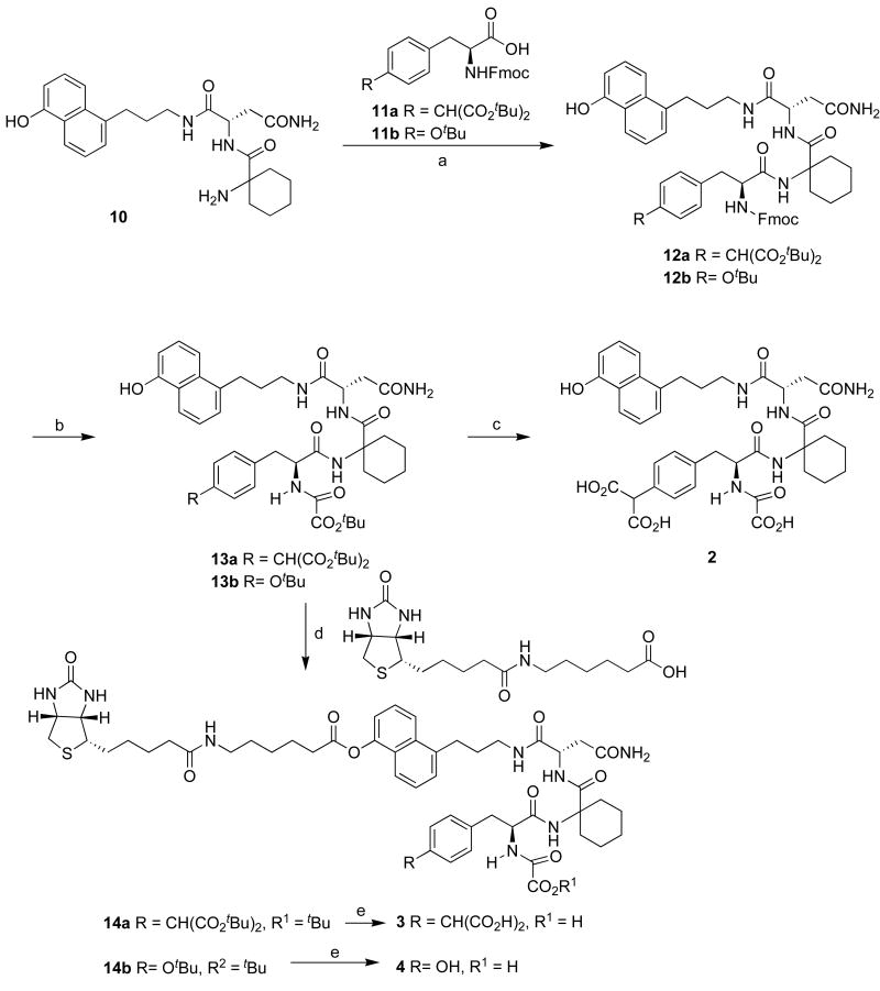
We have shown previously that a potent synthetic antagonist of growth factor receptor-bound protein 2 (Grb2) Src homology 2 (SH2) domain binding (1) blocks growth factor stimulated motility, invasion, and angiogenesis in cultured cell models, as well as tumor metastasis in animals. To characterize the selectivity of 1 for the SH2 domain of Grb2 over other proteins containing similar structural binding motifs, we synthesized a biotinylated derivative (3) that retained high affinity Grb2 SH2 domain binding and potent biological activity. To investigate the selectivity of 1 and 3 for Grb2, the biotinylated antagonist 3 was used to immobilize target proteins from cell extracts for subsequent identification by mass spectrometry. Non-specific binding was identified in parallel using a biotinylated analogue that lacked a single critical binding determinant. The mechanism of action of the antagonist was further characterized by immunoprecipitation, immunoblotting, and light microscopy. This approach to defining protein binding antagonist selectivity and molecular basis of action should be widely applicable in drug development.
Figures
Similar articles
-
Synthesis and structural characterization of a monocarboxylic inhibitor for GRB2 SH2 domain.Bioorg Med Chem Lett. 2021 Nov 1;51:128354. doi: 10.1016/j.bmcl.2021.128354. Epub 2021 Sep 7. Bioorg Med Chem Lett. 2021. PMID: 34506932 Free PMC article.
-
Discovery of a novel nonphosphorylated pentapeptide motif displaying high affinity for Grb2-SH2 domain by the utilization of 3'-substituted tyrosine derivatives.J Med Chem. 2006 Mar 9;49(5):1585-96. doi: 10.1021/jm050910x. J Med Chem. 2006. PMID: 16509576
-
Inhibition of angiogenesis by growth factor receptor bound protein 2-Src homology 2 domain bound antagonists.Mol Cancer Ther. 2004 Oct;3(10):1289-99. Mol Cancer Ther. 2004. PMID: 15486196
-
Grb2 SH2 domain-binding peptide analogs as potential anticancer agents.Biopolymers. 2003;71(2):132-40. doi: 10.1002/bip.10396. Biopolymers. 2003. PMID: 12767115 Review.
-
Structure-based design of compounds inhibiting Grb2-SH2 mediated protein-protein interactions in signal transduction pathways.Curr Pharm Des. 2000 Dec;6(18):1777-96. doi: 10.2174/1381612003398546. Curr Pharm Des. 2000. PMID: 11102562 Review.
Cited by
-
3,4-methylenedioxy-β-nitrostyrene inhibits NLRP3 inflammasome activation by blocking assembly of the inflammasome.J Biol Chem. 2014 Jan 10;289(2):1142-50. doi: 10.1074/jbc.M113.515080. Epub 2013 Nov 21. J Biol Chem. 2014. PMID: 24265316 Free PMC article.
-
GRB2: A dynamic adaptor protein orchestrating cellular signaling in health and disease.Biochem Biophys Rep. 2024 Jul 29;39:101803. doi: 10.1016/j.bbrep.2024.101803. eCollection 2024 Sep. Biochem Biophys Rep. 2024. PMID: 39175664 Free PMC article. Review.
-
Synthesis and structural characterization of a monocarboxylic inhibitor for GRB2 SH2 domain.Bioorg Med Chem Lett. 2021 Nov 1;51:128354. doi: 10.1016/j.bmcl.2021.128354. Epub 2021 Sep 7. Bioorg Med Chem Lett. 2021. PMID: 34506932 Free PMC article.
-
Application of ring-closing metathesis to Grb2 SH3 domain-binding peptides.Biopolymers. 2011;96(6):780-8. doi: 10.1002/bip.21692. Biopolymers. 2011. PMID: 21830199 Free PMC article.
References
-
- Blume-Jensen P, Hunter T. Oncogenic kinase signalling. Nature. 2001;411:355–365. - PubMed
-
- Lowenstein EJ, Daly RJ, Batzer AG, Li W, Margolis B, Lammers R, Ullrich A, Skolnik EY, Bar-Sagi D, Schlessinger J. The SH2 and SH3 domain-containing protein GRB2 links receptor tyrosine kinases to ras signaling. Cell. 1992;70:431–442. - PubMed
-
- Tari AM, Lopez-Berestein G. GRB2: A pivotal protein in signal transduction. Seminars in Oncology. 2001;28:142–147. - PubMed
-
- Kumar R, Gururaj AE, Barnes CJ. p21-activated kinases in cancer. Nat Rev Cancer. 2006;6:459–471. - PubMed
Publication types
MeSH terms
Substances
Grants and funding
LinkOut - more resources
Full Text Sources
Research Materials
Miscellaneous
