

Gallstone ileus following endoscopic retrograde cholangiopancreatography and sphincterotomy: a case report
- PMID: 18990216
- PMCID: PMC2584010
- DOI: 10.1186/1752-1947-2-342
Gallstone ileus following endoscopic retrograde cholangiopancreatography and sphincterotomy: a case report
Abstract
Introduction: Gallstone ileus is a mechanical obstruction caused by the impaction of one or more gallstones within the lumen of any part of the gastrointestinal tract. Although the disorder is a rare cause of small bowel obstruction (1% to 2%), it has been reported to cause up to 25% of cases of non-strangulated small bowel obstruction in patients over 65 years of age.
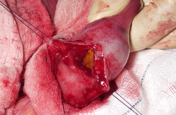
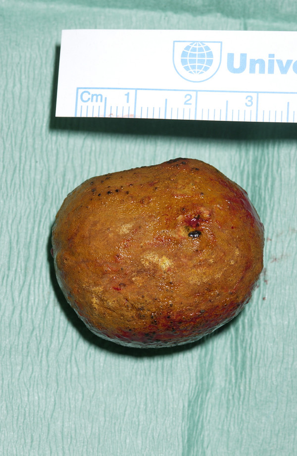
Case presentation: We report a case of a 67-year-old woman who presented with gallstone ileus following endoscopic retrograde cholangiopancreatography and sphincterotomy for choledocholithiasis. She had a history of terminal ileum resection with ileocolic anastomosis for Crohn's disease. A 3 cm gallstone was found to be impacted just proximal to the previous ileocolic anastomosis. A second gallstone was found on digital examination of the proximal small bowel.
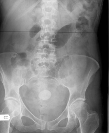
Conclusion: A gallstone may enter the gastrointestinal tract following endoscopic retrograde cholangiopancreatography and sphincterotomy and impact proximal to an anastomotic stricture as demonstrated here. The radiographic image of small bowel obstruction plus air in the biliary tree is a classic diagnostic finding. After stone extraction, the entire small bowel and colon should be digitally examined for further stones.
Figures
References
-
- Reisner RM, Cohen JR. Gallstone ileus: a review of 1001 reported cases. Am Surg. 1994;60:441–446. - PubMed
LinkOut - more resources
Full Text Sources
