

The bovine papillomavirus E5 protein and the PDGF beta receptor: it takes two to tango
- PMID: 18990418
- PMCID: PMC2661243
- DOI: 10.1016/j.virol.2008.09.033
The bovine papillomavirus E5 protein and the PDGF beta receptor: it takes two to tango
Abstract
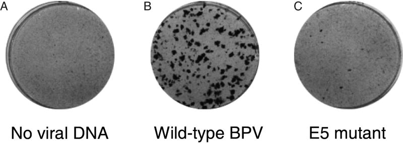
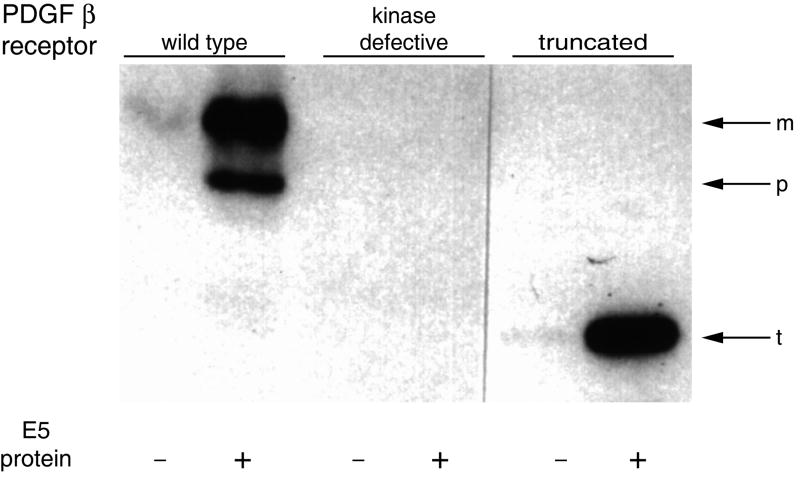
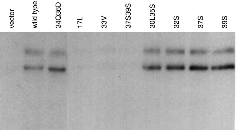
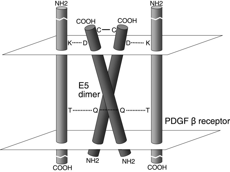
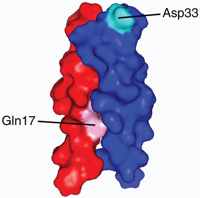
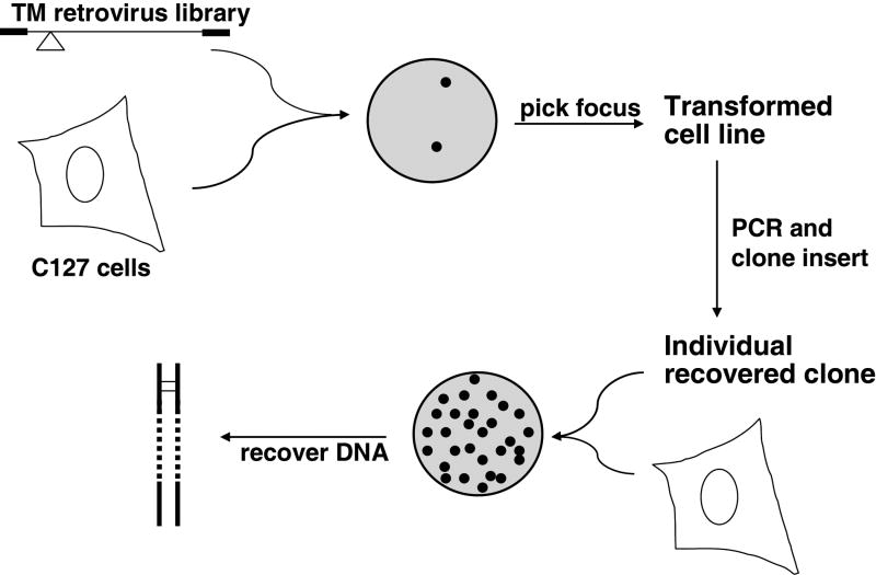
The extremely hydrophobic, 44-amino acid bovine papillomavirus (BPV) E5 protein is the smallest known oncoprotein, which orchestrates cell transformation by causing ligand-independent activation of a cellular receptor tyrosine kinase, the platelet-derived growth factor beta receptor (PDGFbetaR). The E5 protein forms a dimer in transformed cells and is essentially an isolated membrane-spanning segment that binds directly to the transmembrane domain of the PDGFbetaR, inducing receptor dimerization, autophosphorylation, and sustained mitogenic signaling. There are few sequence constraints for activity as long as the overall hydrophobicity of the E5 protein and its ability to dimerize are preserved. Nevertheless, the E5 protein is highly specific for the PDGFbetaR and does not activate other cellular proteins. Genetic screens of thousands of small, artificial hydrophobic proteins with randomized transmembrane domains inserted into an E5 scaffold identified proteins with diverse transmembrane sequences that activate the PDGFbetaR, including some activators as small as 32-amino acids. Analysis of these novel proteins has provided new insight into the requirements for PDGFbetaR activation and specific transmembrane recognition in general. These results suggest that small, transmembrane proteins can be constructed and selected that specifically bind to other cellular or viral transmembrane target proteins. By using this approach, we have isolated a 44-amino acid artificial transmembrane protein that appears to activate the human erythropoietin receptor. Studies of the tiny, hydrophobic BPV E5 protein have not only revealed a novel mechanism of viral oncogenesis, but have also suggested that it may be possible to develop artificial small proteins that specifically modulate much larger target proteins by acting within cellular or viral membranes.
Figures


Similar articles
-
A single amino acid substitution converts a transmembrane protein activator of the platelet-derived growth factor β receptor into an inhibitor.J Biol Chem. 2013 Sep 20;288(38):27273-27286. doi: 10.1074/jbc.M113.470054. Epub 2013 Aug 1. J Biol Chem. 2013. PMID: 23908351 Free PMC article.
-
Two transmembrane dimers of the bovine papillomavirus E5 oncoprotein clamp the PDGF β receptor in an active dimeric conformation.Proc Natl Acad Sci U S A. 2017 Aug 29;114(35):E7262-E7271. doi: 10.1073/pnas.1705622114. Epub 2017 Aug 14. Proc Natl Acad Sci U S A. 2017. PMID: 28808001 Free PMC article.
-
Artificial transmembrane oncoproteins smaller than the bovine papillomavirus E5 protein redefine sequence requirements for activation of the platelet-derived growth factor beta receptor.J Virol. 2009 Oct;83(19):9773-85. doi: 10.1128/JVI.00946-09. Epub 2009 Jul 15. J Virol. 2009. PMID: 19605488 Free PMC article.
-
Mechanisms of cell transformation by papillomavirus E5 proteins.Oncogene. 2001 Nov 26;20(54):7866-73. doi: 10.1038/sj.onc.1204915. Oncogene. 2001. PMID: 11753669 Review.
-
The platelet-derived growth factor beta receptor as a target of the bovine papillomavirus E5 protein.Cytokine Growth Factor Rev. 2000 Dec;11(4):283-93. doi: 10.1016/s1359-6101(00)00012-5. Cytokine Growth Factor Rev. 2000. PMID: 10959076 Review.
Cited by
-
De novo selection of oncogenes.Proc Natl Acad Sci U S A. 2014 Jan 7;111(1):E6-E14. doi: 10.1073/pnas.1315298111. Epub 2013 Dec 16. Proc Natl Acad Sci U S A. 2014. PMID: 24344264 Free PMC article.
-
Role of herpes simplex virus VP11/12 tyrosine-based motifs in binding and activation of the Src family kinase Lck and recruitment of p85, Grb2, and Shc.J Virol. 2013 Oct;87(20):11276-86. doi: 10.1128/JVI.01702-13. Epub 2013 Aug 14. J Virol. 2013. PMID: 23946459 Free PMC article.
-
The HPV16 E6 oncoprotein causes prolonged receptor protein tyrosine kinase signaling and enhances internalization of phosphorylated receptor species.PLoS Pathog. 2013 Mar;9(3):e1003237. doi: 10.1371/journal.ppat.1003237. Epub 2013 Mar 14. PLoS Pathog. 2013. PMID: 23516367 Free PMC article.
-
Cancer associated human papillomaviruses.Curr Opin Virol. 2012 Aug;2(4):459-66. doi: 10.1016/j.coviro.2012.05.004. Epub 2012 Jun 2. Curr Opin Virol. 2012. PMID: 22658985 Free PMC article. Review.
-
Molecular and Phylogenetic Analysis of Bovine Papillomavirus Type 1: First Report in Iraqi Cattle.Adv Virol. 2016;2016:2143024. doi: 10.1155/2016/2143024. Epub 2016 Jun 20. Adv Virol. 2016. PMID: 27413374 Free PMC article.
References
-
- Adduci AJ, Schlegel R. The transmembrane domain of the E5 oncoprotein contains functionally discrete helical faces. J Biol Chem. 1999;274:10249–10258. - PubMed
-
- Andresson T, Sparkowski J, Goldstein DJ, Schlegel R. Vacuolar H+-ATPase mutants transform cells and define a binding site for the papillomavirus E5 oncoprotein. J Biol Chem. 1995;270:6830–6837. - PubMed
-
- Bergman P, Ustav M, Sedman J, Moreno-Lopez J, Vennstrom B, Pettersson U. The E5 gene of bovine papillomavirus type 1 is sufficient for complete oncogenic transformation of mouse fibroblasts. Oncogene. 1988;2:453–459. - PubMed
Publication types
MeSH terms
Substances
Grants and funding
LinkOut - more resources
Full Text Sources
