. 2009 Feb;30(4):649-57.
doi: 10.1016/j.biomaterials.2008.10.003. Epub 2008 Nov 5.

A ligand-mediated nanovector for targeted gene delivery and transfection in cancer cells

Affiliations

A ligand-mediated nanovector for targeted gene delivery and transfection in cancer cells

Omid Veiseh et al. Biomaterials. 2009 Feb.

Abstract

As conventional cancer therapies struggle with toxicity issues and irregular remedial efficacy, the preparation of novel gene therapy vectors could offer clinicians the tools for addressing the genetic errors of diseased tissue. The transfer of gene therapy to the clinic has proven difficult due to safety, target specificity, and transfection efficiency concerns. Polyethylenimine (PEI) nanoparticles have been identified as promising gene carriers that induce gene transfection with high efficiency. However, the inherent toxicity of the material and non-selective delivery are the major concerns in applying these particles clinically. Here, a non-viral nanovector has been developed by PEGylation of DNA-complexing PEI in nanoparticles functionalized with an Alexa Fluor 647 near infrared fluorophore, and the chlorotoxin (CTX) peptide which binds specifically to many forms of cancer. With this nanovector, the potential toxicity to healthy cells is minimized by both the reduction of the toxicity of PEI with the biocompatible copolymer and the targeted delivery of the nanovector to cancer cells, as evaluated by viability studies. The nanovector demonstrated high levels of targeting specificity and gene transfection efficiency with both C6 glioma and DAOY medulloblastoma tumor cells. Significantly, with the CTX as the targeting ligand, the nanovector may serve as a widely applicable gene delivery system for a broad array of cancer types.


Figures

Figure 1
Figure 1
Nanovector preparation scheme. (a) PEGylation of PEI polymer and modification with Alexa Fluor 647 fluorophores (AF). (b) P-PEG-AF modification with SIAX, modification of chlorotoxin (CTX) with traut’s reagent to produce free thiols on peptide and subsequent reaction of the thiol modified CTX peptide with reactive iodoacetate group of P-PEG-AF-SIX yielding P-PEG-AF-CTX. (c) The polymeric construct complexed with DNA to generate the targeting nanovector (P-PEG-AF-CTX/DNA).
Figure 2
Figure 2
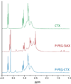
1H NMR spectra showing characteristic peaks of the chlorotoxin peptide (CTX) at 2.60–3.10 and 3.65–3.80 ppm, SIAX-functionalized P-PEG polymer (P-PEG-SIAX) at 3.60–3.65 for the -O-CH2-CH2- repeating unit of PEG and at 2.50–3.10 for the-NH-CH2-CH2- repeating unit of PEI, and the CTX-polymer conjugate (P-PEG-CTX) which shows the characteristic peaks of both CTX and P-PEG.
Figure 3
Figure 3
Agarose gel retardation assay of the polymeric nanovectors made with different polymer:DNA ratios (w:w). (a) Ethidium bromide-stained gel images of DNA-complexed P-PEG-AF-CTX and P-PEG-AF-SIAX nanovectors. (b) Ethidium bromide exclusion assay comparing percent complex formation as a function of polymer:DNA ratio tested for each nanovector.
Figure 4
Figure 4
Zeta potential and hydrodynamic size analysis of nanovectrors. (a) Zeta potentials of nanovectors prepared with varying polymer:DNA ratios (w:w). CTX-modified and unmodified nanovectors shared similar zeta potentials. (b) Hydrodynamic size, by intensity, of nanovectors formed at a polymer:DNA ratio of 5:1 (w:w).
Figure 5
Figure 5
Cell fluorescence intensity analysis by flow cytometry. C6 cells were exposed to either CTX-modified or unmodified nanovectors at varying DNA concentrations.
Figure 6
Figure 6
Flow cytometry based analysis for relative uptake of nanovectors for targeted (C6) and non-targeted (NIH3T3) cells. C6 and NIH3T3 cells were exposed to either the targeting (P-PEG-AF-CTX/DNA) or non-targeting (P-PEG-AF-SIAX/DNA) nanovector at a DNA concentration of 10 µg mL−1.
Figure 7
Figure 7
Analysis of transgene expression of GFP by C6 cells. Transfection efficiency is measured by flow cytometry after treatment with either P-PEG-AF-SIAX/DNA or P-PEG-AF-CTX/DNA at varying DNA concentrations.
Figure 8
Figure 8
Transfection efficiencies of C6 and NIH3T3 cells by DNA complexed with different polymeric vectors, as determined by flow cytometry. The commercially available Lipofectamine was tested as a reference transfection reagent.
Figure 9
Figure 9
Viability analyses of C6 and NIH3T3 cells after exposure to varying transfection vectors. An Alamar blue-based assay was used to determine cell viability.
Figure 10
Figure 10
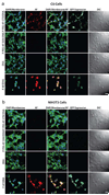
Confocal fluorescence and differential interference contrast (DIC) images of (a) C6 and (b) NIH3T3 cells treated with 10 µg DNA mL−1 without a delivery vector (DNA) or with vectors complexed with either PEI (P-AF/DNA), PEGylated PEI (P-PEG-AF-SIAX/DNA), or PEGylated and CTX-enabled PEI (P-PEG-AF-CTX/DNA). Cellular membranes are shown in green, nuclei in blue, polymeric vectors in red, and GFP expression in turquoise. Scare bars correspond to 40 µm.
Figure 11
Figure 11
Mean levels of transgene expression (mean FITC intensity) in C6, DAOY, and NIH3T3 cells treated with CTX modified (P-PEI-AF-CTX/DNA) or unmodified (P-PEG-AF-SIAX/DNA) nanovectors (5 µg DNA ml−1). As reference the baseline fluorescence intensity of each cell type (Untreated) is also shown.

References

    1. Stewart BW, Kleihues P. World Cancer Report. Geneva: World Health Organization Press; 2003.
    1. Ries L, Melbert D, Krapcho M, Stinchcomb DG, Howlader N, Horner MJ, et al. NCI, editor. SEER Cancer Statistics Review, 1975–2005. Bethesda, MD: 2007. SEER web site http://seer.cancer.gov/csr/1975_2005/
    1. Alton E, Ferrari S, Griesenbach U. Progress and prospects: Gene therapy clinical trials (part 2) Gene Ther. 2007;14(22):1555–1563. - PubMed
    1. Li SD, Huang L. Gene therapy progress and prospects: non-viral gene therapy by systemic delivery. Gene Ther. 2006;13(18):1313–1319. - PubMed
    1. McNeish IA, Bell SJ, Lemoine NR. Gene therapy progress and prospects: cancer gene therapy using tumour suppressor genes. Gene Ther. 2004;11(6):497–503. - PubMed

Publication types

MeSH terms