. 2008 Nov;55(11):2608-19.
doi: 10.1109/TBME.2008.2001294.

Vestibulo-ocular responses evoked via bilateral electrical stimulation of the lateral semicircular canals

Affiliations

Vestibulo-ocular responses evoked via bilateral electrical stimulation of the lateral semicircular canals

Wangsong Gong et al. IEEE Trans Biomed Eng. 2008 Nov.

Abstract

We investigated the vestibulo-ocular responses (VORs) evoked by bilateral electrical stimulation of the nerves innervating horizontal semicircular canals in squirrel monkeys and compared these responses to those evoked by unilateral stimulation. In response to sinusoidal modulation of the electrical pulse rate, the VOR for bilateral stimulation roughly equals the addition of the responses evoked by unilateral right ear and unilateral left ear stimulation; the VOR time constants were about the same for bilateral and unilateral stimulation and both were much shorter than for normal animals. In response to individual pulse stimulation, the VOR evoked by bilateral stimulation closely matches the point-by-point addition of responses evoked by unilateral right ear and unilateral left ear stimulation. We conclude that, to first order, the VOR responses evoked by bilateral stimulation are the summation of the responses evoked by unilateral stimulation. These findings suggest that--from a physiologic viewpoint--unilateral and bilateral vestibular prostheses are about equally viable. Given these findings, one possible advantage of a bilateral prosthesis is higher gain. However, at least for short-term stimulation such as that studied herein, no inherent advantage in terms of the response time constant ("velocity storage") was found.


Figures

Fig. 1
Fig. 1
(A) Horizontal eye responses of monkey “G” evoked by individual biphasic pulse stimulation (300 μA) of the right ear lateral semicircular canal. The evoked responses “piggybacked” on a small (circa 2-3°/s) baseline nystagmus in the dark. (B) Enlargement of content in the small rectangle in (A). Note the background noise and the slope caused by nystagmus. (C) Biphasic current pulses corresponding to (B). (D) The top trace (gray) shows the result of averaging for the data shown in (A) in the -1 degree eye position group. The bottom trace (black) is the response after removal of the slope (slow phase of nystagmus). The vertical line indicates the response peak. (E) A biphasic current pulse corresponding to (D). For eye movement responses, left is positive.
Fig. 2
Fig. 2
Horizontal eye responses of monkey “N” evoked by modulated electrical stimulation and rotation. The top plot in the leftmost column shows the modulation signal (A) that modulated the pulse rate ranging from 133 pps to 267 pps around the baseline of 200 pps, which was equivalent to the modulation provided in our earlier study [5] for a rotation amplitude 80 °/s. The example shown is for 0.1 Hz. The rest of the column shows the eVOR slow phase velocity (SPV) evoked by the modulated continuous electrical stimulation of bilateral push/pull (B), bilateral common mode (C), the right ear (D), and the left ear (E). The second column show the summation (G) and subtraction (H) of the responses to the right and left ear electrical stimulation. The third column shows the responses to yaw rotation (I) when the animal was normal (J) and when both of the horizontal canals were plugged (K). For electrical stimulation, the amplitudes of the current pulses to the right ear and left ear were both set at 125 μA.
Fig. 3
Fig. 3
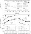
Bode plots of horizontal eye responses of monkey “N”. (A) Gain and (B) phase of horizontal VOR evoked by modulated electrical stimulation of the right ear (▷), the left ear (◁), bilateral push/pull (◊), bilateral common mode (□), the summation (♦) and subtraction (■) of the responses to the right and left ear electrical stimulation, the responses to yaw rotation when the animal was normal (○), and when the canals were plugged (×). Standard error of the mean (SEM) is shown as vertical bars. Phases are not shown for bilateral common mode stimulation, subtraction of unilateral stimulation, and rotation after plugging because the VOR amplitudes were too small to compute phase information reliably.
Fig. 4
Fig. 4
(A) Average horizontal eye responses of monkey “G” evoked by individual biphasic pulses (after slope removal) at different eye positions. The first column (from left) is for the “R only” stimulation, the second column is for the “L only” stimulation. The traces in the third column are the point-to-point addition of the “R only” and the “L only” responses. The fourth column shows responses evoked by “Bilateral” stimulation. In each column, the traces (from bottom to top) show the response average at eye positions from -5 degrees to 0 degree. The spaces between traces in each column are only to separate the traces and do not represent absolute eye position. The traces start 10 ms prior to the current pulse, which is not shown. (B) Peak amplitudes of the traces shown in (A). The responses evoked by bilateral stimulation (◊) match the mathematical addition (♦) of the “R only” (▷) and “L only” (◁) responses.
Fig. 5
Fig. 5
(A) Average horizontal eye response of monkey “G” evoked by individual biphasic pulses (after slope removal) at different current levels designed to yield near-cancellation of the evoked responses. The first column (from left) is for the “R only” stimulation, the second column is for the “L only” stimulation. The traces in the third column are the point-to-point addition of the “R only” and the “L only” responses. The fourth column shows responses evoked by “Bilateral” stimulation. In each column, the traces (from bottom to top) show the response average for current levels from 60 μA to 300 μA for right ear and 30 μA to 110 μA for left ear. The spaces between traces in each column are only to separate the traces and do not represent absolute eye position. The traces start 10 ms prior to the current pulse. (B) Peak amplitudes of the traces shown in (A). The responses evoked by bilateral stimulation (◊) match the mathematical addition (♦) of the “R only” (▷) and “L only” (◁) responses.
Fig. 6
Fig. 6
(A) Average horizontal eye response of monkey “G” evoked by individual biphasic pulse (after slope removal). The first column (from left) is for the “R only” stimulation, the second column is for the “L only” stimulation. The traces in the third column are the point-to-point addition of the “R only” and the “L only” responses. The fourth column shows responses evoked by “Bilateral” stimulation. In each column, the traces (from bottom to top) show the response average for current levels from 60 μA to 300 μA for the right ear and 70 μA for the left ear. The spaces between traces in each column are only to separate the traces and do not represent absolute eye position. The traces start 10 ms prior to the current pulse. (B) Peak amplitudes of the traces shown in (A). The responses evoked by bilateral stimulation (◊) match the mathematical addition (♦) of the “R only” (▷) and “L only” (◁) responses. (C) Peak amplitudes of the responses evoked by individual biphasic pulse when the current level applied to the right ear was fixed at 180 μA while the current to the left ear varied between 30 μA and 110 μA. The responses evoked by bilateral stimulation (◊) match the mathematical addition (♦) of the “R only” and “L only” (◁) responses.
Fig. 7
Fig. 7
(A) Average horizontal eye response of monkey “G” evoked by individual biphasic pulse (after slope removal) with the current pulse to the right ear delayed relative to the left ear stimulation. The first column (from left) is the response to the right ear at 300 μA, the second column is the response to the left ear at 110 μA. The traces in the third column show the point-to-point addition of the “R only” and the “L only” responses when the response to the “R only” was shifted in time by 0, 0.6, 1, 1.5, 2, 2.5, and 3 ms (for traces from top to bottom). The fourth column shows the responses evoked by “Bilateral” stimulation. The traces (from top to bottom) are the responses to the stimulation pulse with the pulse to the right ear delayed 0, 0.6, 1, 1.5, 2, 2.5, 3 ms. Spaces between traces in each column only separate the traces and do not represent absolute eye position. The traces start 10 ms prior to the current pulse to the left ear. (B) Peak amplitude of the traces shown in (A). The responses evoked by bilateral stimulation (◊) match the mathematical addition (♦) of the “R only” and “L only” responses. (C) Peak amplitudes of the responses when the current to the left ear was delayed. The responses evoked by bilateral stimulation (◊) match the mathematical addition (♦) of the “R only” and “L only” responses.
Fig. 8
Fig. 8
(A) Average horizontal eye response of monkey “G” to individual pulses to the right ear at current levels (from bottom to top) from 96 μA to 320 μA for anodic and cathodic pulses and from 90 μA to 300 μA for biphasic pulses. (B) Peak amplitudes of the responses evoked by right ear stimulation via anodic ( formula image), cathodic (▶), and biphasic (▷) pulses. The responses evoked by cathodic and biphasic pulses are indistinguishable, while those evoked by anodic pulses are smaller. (C) Average horizontal eye response to individual pulses applied to the left ear at current levels (from bottom to top) from 40 μA to 110 μA for anodic, cathodic, and biphasic pulses. (D) Peak amplitude of responses evoked by left ear stimulation via anodic ( formula image), cathodic (◀), and biphasic (◁) pulses. The responses evoked by cathodic and biphasic pulses are indistinguishable, while those evoked by anodic pulses are smaller.

References

    1. Gong W, Merfeld D. A prototype neural semicircular canal prosthesis using patterned electrical stimulation. Ann Biomed Eng. 2000;28:572–581. - PubMed
    1. Gong W, Merfeld D. System design and performance of a unilateral semicircular canal prosthesis. IEEE Trans Biomed Eng. 2002;49:175–181. - PubMed
    1. Merfeld D, Gong W, Morrissey J, Saginaw M, Haburcakova C, Lewis R. Acclimation to chronic constant-rate peripheral stimulation provided by a vestibular prosthesis. IEEE Trans Biomed Eng. 2006;53:2362–2372. - PubMed
    1. Lewis RF, Gong W, Ramsey M, Minor L, Boyle R, Merfeld DM. Vestibular adaptation studied with a prosthetic semicircular canal. J Vestib Res. 2002;12:87–94. - PubMed
    1. Merfeld D, Haburcakova C, Gong W, Lewis R. Chronic Vestibulo-ocular Reflexes Evoked by a Vestibular Prosthesis. IEEE Trans Biomed Eng. 2007;54:1005–1015. - PubMed

Publication types