

Identification and purification of mesodermal progenitor cells from human adult bone marrow
- PMID: 18991503
- PMCID: PMC3085824
- DOI: 10.1089/scd.2008.0291
Identification and purification of mesodermal progenitor cells from human adult bone marrow
Abstract
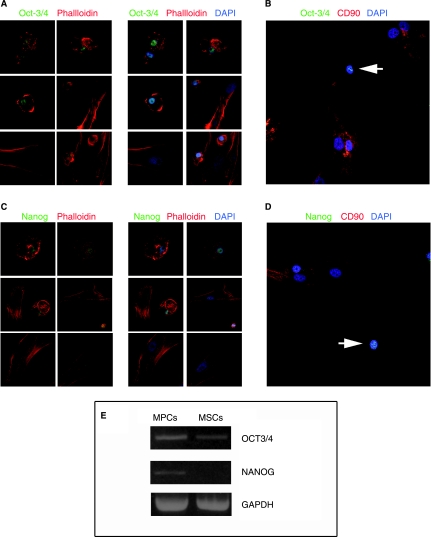
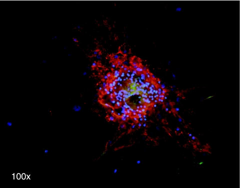
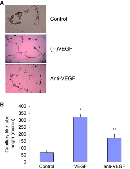
Bone marrow-derived mesodermal stem cells may differentiate toward several lines and are easily cultured in vitro. Some putative progenitors of these cells have been described in both humans and mice. Here, we describe a new mesodermal progenitor population [mesodermal progenitors cells (MPCs)] able to differentiate into mesenchymal cells upon appropriate culture conditions. When cultured in presence of autologous serum, these cells are strongly adherent to plastic, resistant to trypsin detachment, and resting. Mesodermal progenitor cells may be pulsed to proliferate and differentiate by substituting autologous serum for human cord blood serum or fetal calf serum. By these methods cells proliferate and differentiate toward mesenchymal cells and thus may further differentiate into osteoblats, chondrocytes, or adipocytes. Moreover MPCs are capable to differentiate in endothelial cells (ECs) showing characteristics similar to microvessel endothelium cells. Mesodermal progenitors cells have a defined phenotype and carry embryonic markers not present in mesenchymal cells. Moreover MPCs strongly express aldehyde dehydrogenase activity, usually present in hematopoietic precursors but absent in mesenchymal cells. When these progenitors are pulsed to differentiate, they lose these markers and acquire the mesenchymal ones. Interestingly, mesenchymal cells may not be induced to back differentiate into MPCs. Our results demonstrate the adult serum role in maintaining pluripotent mesodermal precursors and allow isolation of these cells. After purification, MPCs may be pulsed to proliferate in a very large scale and then induced to differentiate, thus possibly allowing their use in regenerative medicine.
Figures
Similar articles
-
Selective culture of mesodermal progenitor cells.Stem Cells Dev. 2009 Oct;18(8):1227-34. doi: 10.1089/scd.2009.0054. Stem Cells Dev. 2009. PMID: 19331526
-
Purification and ex vivo expansion of postnatal human marrow mesodermal progenitor cells.Blood. 2001 Nov 1;98(9):2615-25. doi: 10.1182/blood.v98.9.2615. Blood. 2001. Retraction in: Blood. 2009 Mar 5;113(10):2370. doi: 10.1182/blood-2008-10-186288. PMID: 11675329 Retracted.
-
Mesangiogenic Progenitor Cells Derived from One Novel CD64(bright)CD31(bright)CD14(neg) Population in Human Adult Bone Marrow.Stem Cells Dev. 2016 May 1;25(9):661-73. doi: 10.1089/scd.2015.0344. Epub 2016 Apr 11. Stem Cells Dev. 2016. PMID: 26975798 Free PMC article.
-
Mesenchymal stem cells.Exp Biol Med (Maywood). 2001 Jun;226(6):507-20. doi: 10.1177/153537020122600603. Exp Biol Med (Maywood). 2001. PMID: 11395921 Review.
-
Trafficking and differentiation of mesenchymal stem cells.J Cell Biochem. 2009 Apr 15;106(6):984-91. doi: 10.1002/jcb.22091. J Cell Biochem. 2009. PMID: 19229871 Review.
Cited by
-
WNT5B in Physiology and Disease.Front Cell Dev Biol. 2021 May 4;9:667581. doi: 10.3389/fcell.2021.667581. eCollection 2021. Front Cell Dev Biol. 2021. PMID: 34017835 Free PMC article. Review.
-
Targeting of stromal versican by miR-144/199 inhibits multiple myeloma by downregulating FAK/STAT3 signalling.RNA Biol. 2020 Jan;17(1):98-111. doi: 10.1080/15476286.2019.1669405. Epub 2019 Sep 29. RNA Biol. 2020. PMID: 31532704 Free PMC article.
-
Are MSCs angiogenic cells? New insights on human nestin-positive bone marrow-derived multipotent cells.Front Cell Dev Biol. 2014 May 20;2:20. doi: 10.3389/fcell.2014.00020. eCollection 2014. Front Cell Dev Biol. 2014. PMID: 25364727 Free PMC article.
-
Constitutive expression of pluripotency-associated genes in mesodermal progenitor cells (MPCs).PLoS One. 2010 Mar 25;5(3):e9861. doi: 10.1371/journal.pone.0009861. PLoS One. 2010. PMID: 20360837 Free PMC article.
-
Mesangiogenic progenitor cells are forced toward the angiogenic fate, in multiple myeloma.Oncotarget. 2019 Nov 26;10(63):6781-6790. doi: 10.18632/oncotarget.27285. eCollection 2019 Nov 26. Oncotarget. 2019. PMID: 31827721 Free PMC article.
References
-
- Bruder SP. Jaiswal N. Haynesworth SE. Growth kinetics, self-renewal, and the osteogenic potential of purified human mesenchymal stem cells during extensive subcultivation and following cryopreservation. J Cell Biochem. 1997;64:278–294. - PubMed
-
- Caplan AI. Mesenchymal stem cells. J Orthop Res. 1991;9:641–650. - PubMed
-
- Pittenger MF. Mackay AM. Beck SC. Jaiswal RK. Douglas R. Mosca JD. Moorman MA. Simonetti DW. Craig S. Marshak DR. Multilineage potential of adult human mesenchymal stem cells. Science. 1999;284:143–147. - PubMed
-
- Woodbury D. Schwarz EJ. Prockop DJ. Black IB. Adult rat and human bone marrow stromal cells differentiate into neurons. J Neurosci Res. 2000;61:364–370. - PubMed
-
- Barry FP. Boynton RE. Haynesworth S. Murphy JM. Zaia J. The monoclonal antibody SH-2, raised against human mesenchymal stem cells, recognizes an epitope on endoglin (CD105) Biochem Biophys Res Commun. 1999;265:134–139. - PubMed
MeSH terms
Substances
Grants and funding
LinkOut - more resources
Full Text Sources
Other Literature Sources
Medical
