

Neuroimaging in human MDMA (Ecstasy) users
- PMID: 18991874
- PMCID: PMC2677829
- DOI: 10.1196/annals.1432.007
Neuroimaging in human MDMA (Ecstasy) users
Abstract
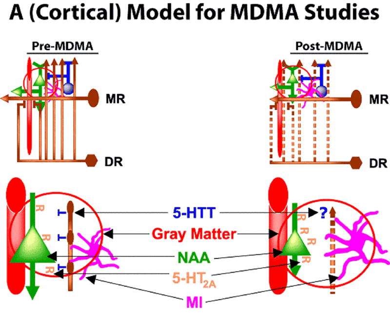
MDMA (3,4 methylenedioxymethamphetamine) has been used by millions of people worldwide as a recreational drug. The terms "MDMA" and "Ecstasy" are often used synonymously, but it is important to note that the purity of Ecstasy sold as MDMA is not certain. MDMA use is of public health concern, not so much because MDMA produces a common or severe dependence syndrome, but rather because rodent and nonhuman primate studies have indicated that MDMA (when administered at certain dosages and intervals) can cause long-lasting reductions in markers of brain serotonin (5-HT) that appear specific to fine-diameter axons arising largely from the dorsal raphe nucleus (DR). Given the popularity of MDMA, the potential for the drug to produce long-lasting or permanent 5-HT axon damage or loss, and the widespread role of 5-HT function in the brain, there is a great need for a better understanding of brain function in human users of this drug. To this end, neuropsychological, neuroendocrine, and neuroimaging studies have all suggested that human MDMA users may have long-lasting changes in brain function consistent with 5-HT toxicity. Data from animal models leads to testable hypotheses regarding MDMA's effects on the human brain. Because neuropsychological and neuroimaging findings have focused on the neocortex, a cortical model is developed to provide a context for designing and interpreting neuroimaging studies in MDMA users. Aspects of the model are supported by the available neuroimaging data, but there are controversial findings in some areas and most findings have not been replicated across different laboratories and using different modalities. This paper reviews existing findings in the context of a cortical model and suggests directions for future research.
Figures
Similar articles
-
Neuroimaging research in human MDMA users: a review.Psychopharmacology (Berl). 2007 Jan;189(4):539-56. doi: 10.1007/s00213-006-0467-3. Epub 2006 Jul 18. Psychopharmacology (Berl). 2007. PMID: 16847678 Review.
-
Toxicodynamics and long-term toxicity of the recreational drug, 3, 4-methylenedioxymethamphetamine (MDMA, 'Ecstasy').Toxicol Lett. 2000 Mar 15;112-113:143-6. doi: 10.1016/s0378-4274(99)00216-7. Toxicol Lett. 2000. PMID: 10720723 Review.
-
The effects of ecstasy on neurotransmitter systems: a review on the findings of molecular imaging studies.Psychopharmacology (Berl). 2016 Oct;233(19-20):3473-501. doi: 10.1007/s00213-016-4396-5. Epub 2016 Aug 28. Psychopharmacology (Berl). 2016. PMID: 27568200 Free PMC article. Review.
-
(+/-)3,4-Methylenedioxymethamphetamine ('Ecstasy')-induced serotonin neurotoxicity: studies in animals.Neuropsychobiology. 2000;42(1):5-10. doi: 10.1159/000026664. Neuropsychobiology. 2000. PMID: 10867550 Review.
-
Activation of protein kinase C (PKC) by 3,4-methylenedioxymethamphetamine (MDMA) occurs through the stimulation of serotonin receptors and transporter.Neuropsychopharmacology. 1997 Sep;17(3):117-29. doi: 10.1016/S0893-133X(97)00026-2. Neuropsychopharmacology. 1997. PMID: 9272479
Cited by
-
N-acetylaspartate (NAA) correlates inversely with cannabis use in a frontal language processing region of neocortex in MDMA (Ecstasy) polydrug users: a 3 T magnetic resonance spectroscopy study.Pharmacol Biochem Behav. 2009 Mar;92(1):105-10. doi: 10.1016/j.pbb.2008.10.022. Epub 2008 Nov 13. Pharmacol Biochem Behav. 2009. PMID: 19032963 Free PMC article.
-
Evidence for chronically altered serotonin function in the cerebral cortex of female 3,4-methylenedioxymethamphetamine polydrug users.Arch Gen Psychiatry. 2012 Apr;69(4):399-409. doi: 10.1001/archgenpsychiatry.2011.156. Epub 2011 Dec 5. Arch Gen Psychiatry. 2012. PMID: 22147810 Free PMC article.
-
Prior MDMA (Ecstasy) use is associated with increased basal ganglia-thalamocortical circuit activation during motor task performance in humans: an fMRI study.Neuroimage. 2009 Jul 1;46(3):817-26. doi: 10.1016/j.neuroimage.2009.02.029. Epub 2009 Mar 2. Neuroimage. 2009. PMID: 19264142 Free PMC article.
-
3,4-methylenedioxymethamphetamine (MDMA): current perspectives.Subst Abuse Rehabil. 2013 Nov 21;4:83-99. doi: 10.2147/SAR.S37258. eCollection 2013. Subst Abuse Rehabil. 2013. PMID: 24648791 Free PMC article. Review.
-
Human ecstasy (MDMA) polydrug users have altered brain activation during semantic processing.Psychopharmacology (Berl). 2013 May;227(1):41-54. doi: 10.1007/s00213-012-2936-1. Epub 2012 Dec 16. Psychopharmacology (Berl). 2013. PMID: 23241648 Free PMC article.
References
-
- Cole JC, et al. The content of ecstasy tablets: implications for the study of their long-term effects. Addiction. 2002;97:1531–1536. - PubMed
-
- Parrott AC. Is ecstasy MDMA? A review of the proportion of ecstasy tablets containing MDMA, their dosage levels, and the changing perceptions of purity. Psychopharmacology (Berl) 2004;173:234–241. - PubMed
-
- Tanner-Smith EE. Pharmacological content of tablets sold as “ecstasy”: Results from an online testing service. Drug Alcohol Depend. 2006;83:247–254. - PubMed
-
- Green AR, et al. The pharmacology and clinical pharmacology of 3,4-methylenedioxymethamphetamine (MDMA, “ecstasy”) Pharmacol Rev. 2003;55:463–508. - PubMed
-
- Lyles J, Cadet JL. Methylenedioxymethamphetamine (MDMA, Ecstasy) neurotoxicity: cellular and molecular mechanisms. Brain Res Rev. 2003;42:155–168. - PubMed
Publication types
MeSH terms
Substances
Grants and funding
LinkOut - more resources
Full Text Sources
Medical
