Transient TWEAK overexpression leads to a general salivary epithelial cell proliferation
- PMID: 18992019
- PMCID: PMC2760474
- DOI: 10.1111/j.1601-0825.2008.01474.x
Transient TWEAK overexpression leads to a general salivary epithelial cell proliferation
Abstract
Objectives: Tumor necrosis factor-like weak inducer of apoptosis (TWEAK) is a multifunctional cytokine that has pro-apoptotic, pro-angiogenic and pro-inflammatory effects. In liver, TWEAK leads to proliferation of progenitor oval cells, but not of mature hepatocytes. This study evaluated the hypothesis that TWEAK overexpression in salivary glands would lead to the proliferation of a salivary progenitor cell.
Methods: A recombinant, serotype 5 adenoviral vector encoding human TWEAK, AdhTWEAK, was constructed, initially tested in vitro, and then administered to male Balb/c mice via cannulation of Wharton's duct. TWEAK expression in vivo was monitored as protein secreted into saliva and serum by enzyme-linked immunosorbent assays. Salivary cell proliferation was monitored by proliferating cell nuclear antigen staining and apoptosis was monitored using TUNEL staining.
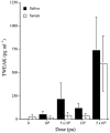
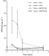
Results: AdhTWEAK administration led to a dose-dependent, transient TWEAK protein expression, detected primarily in saliva. Salivary epithelial cell proliferation was generalized, peaking on approximately days 2 and 3. TWEAK expression had no detectable effect on apoptosis of salivary epithelial cells.
Conclusion: Transient overexpression of TWEAK in murine salivary glands leads to a general proliferation of epithelial cells vs a selective stimulation of a salivary progenitor cell.
Figures


Similar articles
-
TWEAK induces liver progenitor cell proliferation.J Clin Invest. 2005 Sep;115(9):2330-40. doi: 10.1172/JCI23486. Epub 2005 Aug 18. J Clin Invest. 2005. PMID: 16110324 Free PMC article.
-
Gene transfer mediated by different viral vectors following direct cannulation of mouse submandibular salivary glands.Eur J Oral Sci. 2002 Jun;110(3):254-60. doi: 10.1034/j.1600-0722.2002.21200.x. Eur J Oral Sci. 2002. PMID: 12120712
-
The cytokine tumor necrosis factor-like weak inducer of apoptosis and its receptor fibroblast growth factor-inducible 14 have a neuroprotective effect in the central nervous system.J Neuroinflammation. 2012 Mar 6;9:45. doi: 10.1186/1742-2094-9-45. J Neuroinflammation. 2012. PMID: 22394384 Free PMC article.
-
TWEAK/Fn14 signaling in tumors.Tumour Biol. 2017 Jun;39(6):1010428317714624. doi: 10.1177/1010428317714624. Tumour Biol. 2017. PMID: 28639899 Review.
-
TWEAK/Fn14 pathway: an immunological switch for shaping tissue responses.Immunol Rev. 2011 Nov;244(1):99-114. doi: 10.1111/j.1600-065X.2011.01054.x. Immunol Rev. 2011. PMID: 22017434 Review.
Cited by
-
Tumor necrosis factor-like weak inducer of apoptosis and its potential roles in lupus nephritis.Inflamm Res. 2012 Apr;61(4):277-84. doi: 10.1007/s00011-011-0420-8. Inflamm Res. 2012. PMID: 22297307 Review.
-
Evaluation of a rapamycin-regulated serotype 2 adeno-associated viral vector in macaque parotid glands.Oral Dis. 2010 Apr;16(3):269-77. doi: 10.1111/j.1601-0825.2009.01631.x. Oral Dis. 2010. PMID: 20374510 Free PMC article.
-
Transgenic α-1-antitrypsin secreted into the bloodstream from salivary glands is biologically active.Oral Dis. 2011 Jul;17(5):476-83. doi: 10.1111/j.1601-0825.2010.01775.x. Epub 2010 Dec 2. Oral Dis. 2011. PMID: 21122036 Free PMC article.
References
-
- Adesanya MR, Redman RS, Baum BJ, et al. Immediate inflammatory responses to adenovirus-mediated gene transfer in rat salivary glands. Hum Gene Ther. 1996;7:1085–1093. - PubMed
-
- Baum BJ. Principles of saliva secretion. Ann NY Acad Sci. 1993;694:17–23. - PubMed
-
- Baum BJ, Wang S, Cukierman E, et al. Re-engineering the functions of a terminally differentiated cell in vivo. Ann NY Acad Sci. 1999;875:294–300. - PubMed
-
- Baum BJ, Wellner RB, Zheng C. Gene transfer to salivary glands. Int Rev Cytol. 2002;213:93–146. - PubMed
-
- Brown AM, Rusnock EJ, Sciubba JJ, et al. Establishment and characterization of an epithelial cell line from rat submandibular gland. J Oral Pathol Med. 1989;18:206–213. - PubMed
Publication types
MeSH terms
Substances
Grants and funding
LinkOut - more resources
Full Text Sources

