. 2008 Nov 7:8:325.
doi: 10.1186/1471-2407-8-325.

Hepatocyte growth factor enhances death receptor-induced apoptosis by up-regulating DR5

Affiliations

Hepatocyte growth factor enhances death receptor-induced apoptosis by up-regulating DR5

Yang Li et al. BMC Cancer. .

Abstract

Background: Hepatocyte growth factor (HGF) and its receptor c-MET are commonly expressed in malignant gliomas and embryonic neuroectodermal tumors including medulloblastoma and appear to play an important role in the growth and dissemination of these malignancies. Dependent on cell context and the involvement of specific downstream effectors, both pro- and anti-apoptotic effects of HGF have been reported.

Methods: Human medulloblastoma cells were treated with HGF for 24-72 hours followed by death receptor ligand TRAIL (Tumor necrosis factor-related apoptosis-inducing ligand) for 24 hours. Cell death was measured by MTT and Annexin-V/PI flow cytometric analysis. Changes in expression levels of targets of interest were measured by Northern blot analysis, quantitative reverse transcription-PCR, Western blot analysis as well as immunoprecipitation.

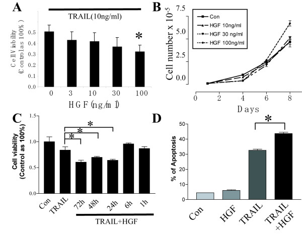
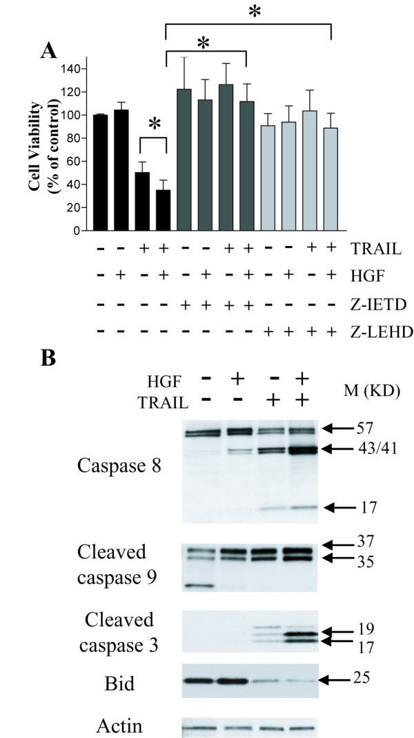
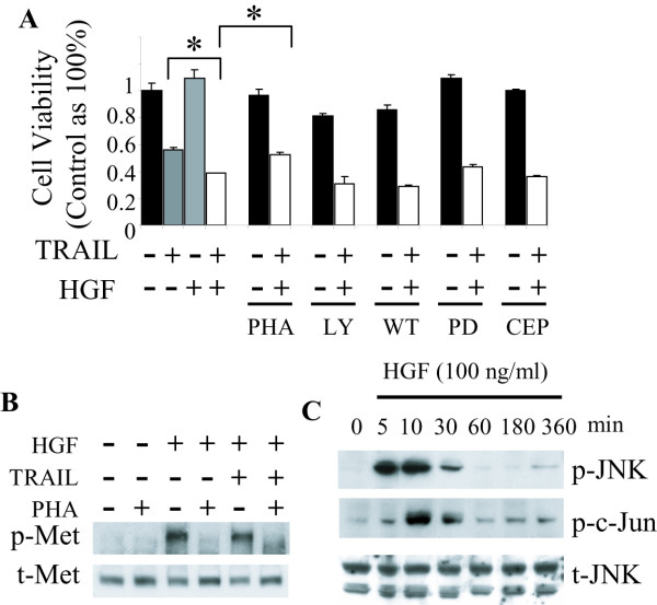
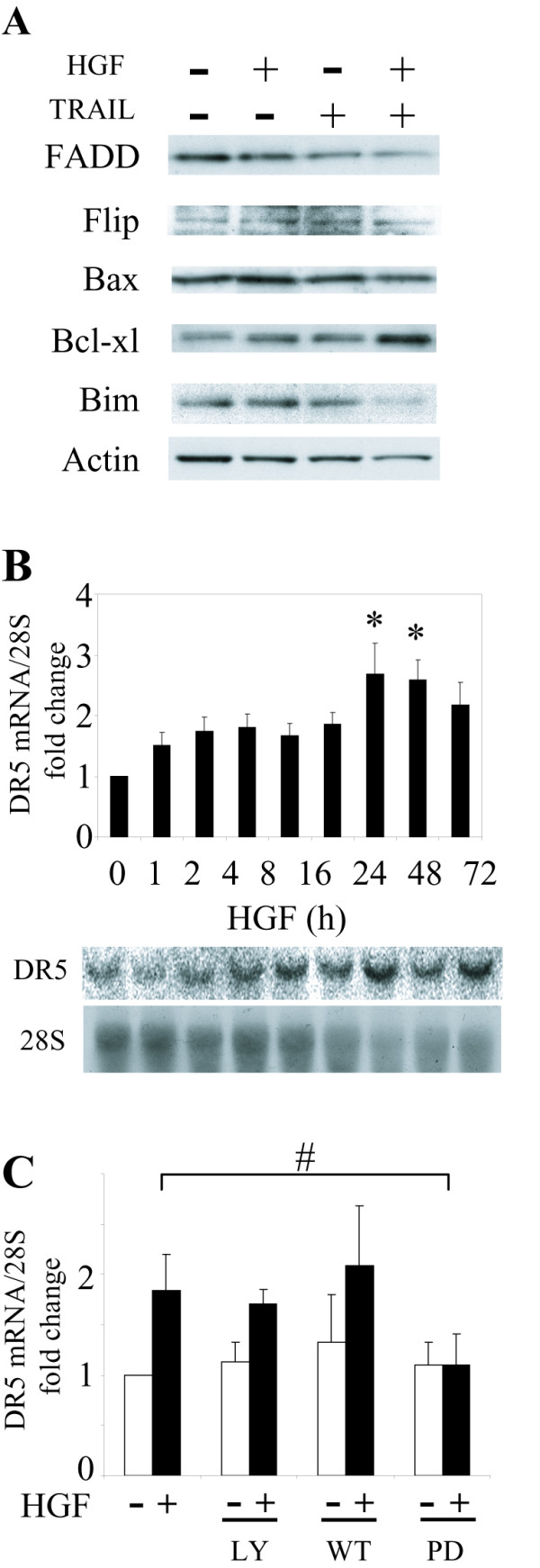
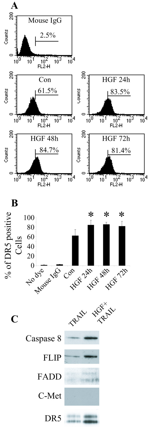
Results: In this study, we show that HGF promotes medulloblastoma cell death induced by TRAIL. TRAIL alone triggered apoptosis in DAOY cells and death was enhanced by pre-treating the cells with HGF for 24-72 h prior to the addition of TRAIL. HGF (100 ng/ml) enhanced TRAIL (10 ng/ml) induced cell death by 36% (P<0.001). No cell death was associated with HGF alone. Treating cells with PHA-665752, a specific c-Met receptor tyrosine kinase inhibitor, significantly abrogated the enhancement of TRAIL-induced cell death by HGF, indicating that its death promoting effect requires activation of its canonical receptor tyrosine kinase. Cell death induced by TRAIL+HGF was predominately apoptotic involving both extrinsic and intrinsic pathways as evidenced by the increased activation of caspase-3, 8, 9. Promotion of apoptosis by HGF occurred via the increased expression of the death receptor DR5 and enhanced formation of death-inducing signal complexes (DISC).

Conclusion: Taken together, these and previous findings indicate that HGF:c-Met pathway either promotes or inhibits medulloblastoma cell death via pathway and context specific mechanisms.


Figures

Figure 1
Figure 1
HGF potentiates TRAIL induced apoptosis in DAOY cells. A. HGF enhances TRAIL induced cell death in a dose-dependent manner as measured by MTT assay. TRAIL at 10 ng/ml induced 50% cell loss. Pre-incubation with HGF (100 ng/ml) for 72 h resulted in 18% more cell death compare to TRAIL alone. B. HGF alone induces DAOY cell growth. Cells were counted every two days after plating with or without HGF in medium containing 10% FBS. HGF promoted cell growth in a dose-dependent manner. C. HGF sensitizes TRAIL (3 ng/ml) induced cell death in a time-dependent manner. Pre-incubation with HGF for at least 24 h was required to enhance TRAIL-induced cell death. D. FACscan analysis of DAOY cell apoptosis using Annexin V-FITC and propidium iodide (PI) staining. Annexin V-positive cells were found to be 6%, 32% and 44% in the presence of HGF (100 ng/ml), TRAIL (10 ng/ml) and HGF+TRAIL, respectively. Experiments were repeated three times. Data represents mean ± SE (N = 9, * P < 0.001).
Figure 2
Figure 2
Involvement of intrinsic and extrinsic apoptotic pathways in cell death induced by TRAIL + HGF. A: MTT assay showing cytoprotection from specific inhibitors of caspase-8 (Z-IETD, 50 μM) and caspase-9 (Z-LEHD, 50 μM) against TRAIL + HGF induced apoptosis. DAOY cells were pre-incubated with HGF for 72 h and then treated with caspase inhibitors for 30 min before adding TRAIL. Z-IETD reversed cell death induced by TRAIL + HGF completely, whereas Z-LEHD protected ~80% of the cells from death. B. Western blot analysis of caspase-8, -9, -3 and Bid. Cells were treated with or without HGF (100 ng/ml) for 72 h before incubation with TRAIL (10 ng/ml) for another 24 h. TRAIL alone induced activation (cleavage) of caspase-3, 8, 9 and Bid. HGF enhanced the activation of caspases and Bid by TRAIL. Experiments were repeated three times (N = 9, * P < 0.001).
Figure 3
Figure 3
Signaling pathways mediating the potentiation of TRAIL-induced cell death by HGF. A. DAOY cells were pre-incubated with or without inhibitors for 30 min, and then treated with HGF for 72 h followed by TRAIL for 24 h. Cell viability was analyzed by MTT assay. The c-Met inhibitor PHA-665752 (100 nM) significantly reduced the cell death promoting effect of HGF. The PI3 kinase inhibitor LY294002 (30 μM) or wortmannin (1 μM) slightly increased the cell death rate induced by TRAIL + HGF. The ERK inhibitor PD98059 (30 μM) as well as the JNK pathway inhibitor CEP-11004 (10 μM) had no effect on the cell death induced by TRAIL + HGF. B. DAOY cells were pre-incubated with c-Met inhibitor PHA-665752 (100 nM) for 30 min before adding HGF. Phosphorylation of c-Met in cells treated with HGF or HGF+TRAIL was inhibited by PHA-665752. C. Western blot analysis with anti-JNK and anti-phospho-JNK (Ser 198) antibodies was used to determine JNK activation. In DAOY cells, HGF induced JNK activation as early as 5 min and lasted for 30 min. C-Jun was also activated within 10 min of HGF treatment. Data represent mean ± SE (N = 9, * P < 0.001).
Figure 4
Figure 4
Modulation of pro- and anti-apoptotic proteins by HGF. A. Western blot analysis of FADD, FLIP, Bax, Bcl-xl and Bim protein levels in response to HGF, TRAIL and HGF + TRAIL. Cells were treated with HGF for 72 h before incubation with TRAIL for another 24 h. FADD and Bim were slightly down regulated by HGF + TRAIL. B. Northern blot analysis of DR5 mRNA levels in response to HGF. DAOY cells were treated with HGF for the indicated times. Total cell RNA was hybridized with probes for DR5 and 28S RNA (control) as described in Materials and Methods. DR5 isoform 1 mRNA levels were increased between 1–72 h as shown (* P < 0.001). C. DAOY cells were pre-incubated with inhibitors for 30 min, and then treated with HGF for 24 h followed by Northern blot analysis for DR5 expression. The PI3K inhibitor LY294002 (30 μM) or wortmannin (1 μM) did not affect the DR5 up-regulation induced by HGF. The MAPK inhibitor PD98059 (30 μM) reversed up-regulation of DR5 by HGF (# P < 0.05). Data represent mean ± SE (N = 6).
Figure 5
Figure 5
HGF increases the DR5 expression and enhances TRAIL-induced DISC formation. A. FACScan analysis of DR5 expression in DAOY cell surface after HGF treatment. Cells treated with HGF for 24 – 72 h were incubated with PE-conjugated anti-DR5 antibody for 30 min. Mouse IgG was used as negative control. At baseline ~61.5% cells were DR5 positive. After 24 h incubation with HGF, the DR5 positive pool increased to 83.5%. B. Compared to control, there was a significant increase in DR5 positive cells after 24 – 72 h incubation with HGF (N = 6, * P < 0.05). C. DAOY cells were pre-treated with HGF for 72 h followed by TRAIL for 2 h. Equal amounts of total cell protein (500 μg) were immunoprecipitated with anti-DR5 antibody and then subjected to Western blot analysis with antibodies to caspase 8, FLIP, FADD, c-Met and DR5 antibody. There was a distinct increased in DISC in cells treated with TRAIL + HGF.
Figure 6
Figure 6
Relationship between c-Met and DR5 expression in human embryonal CNS tumors. A. Quantitative reverse transcription-PCR results showing the expression levels of c-Met and DR5 relative to actin in 18 snap-frozen tissues samples. The expression of c-Met and DR5 in all samples was calculated in relation to DAOY expression. B. We assigned tumors to either high (upper third), median (middle third) and low (lowest third) c-Met expression or TRAIL expression (N = 6 per group). Of the 6 tumors expressing high c-Met (H-cMet), 50% expressed high DR5. Of the 6 tumors expressing low c-Met (L-cMet), 50% expressed low level of DR5. The correlation between c-Met expression and DR5 expression is 0.4 (P = 0.06, Log Pearson analysis).

Similar articles

Cited by

References

    1. Birchmeier C, Birchmeier W, Gherardi E, Woude GF Vande. Met, metastasis, motility and more. Nat Rev Mol Cell Biol. 2003;4:915–925. doi: 10.1038/nrm1261. - DOI - PubMed
    1. Matsumoto K, Nakamura T. Emerging multipotent aspects of hepatocyte growth factor. J Biochem. 1996;119:591–600. - PubMed
    1. Rosen EM, Lamszus K, Laterra J, Polverini PJ, Rubin JS, Goldberg ID. HGF/SF in angiogenesis. Ciba Found Symp. 1997;212:215–226. discussion 227-219. - PubMed
    1. Bottaro DP, Rubin JS, Faletto DL, Chan AM, Kmiecik TE, Woude GF Vande, Aaronson SA. Identification of the hepatocyte growth factor receptor as the c-met proto-oncogene product. Science. 1991;251:802–804. doi: 10.1126/science.1846706. - DOI - PubMed
    1. Rosen EM, Laterra J, Joseph A, Jin L, Fuchs A, Way D, Witte M, Weinand M, Goldberg ID. Scatter factor expression and regulation in human glial tumors. Int J Cancer. 1996;67:248–255. doi: 10.1002/(SICI)1097-0215(19960717)67:2<248::AID-IJC16>3.0.CO;2-7. - DOI - PubMed

Publication types

MeSH terms

Substances