. 2008 Nov 7:8:106.
doi: 10.1186/1471-213X-8-106.

Expression of BLIMP1/PRMT5 and concurrent histone H2A/H4 arginine 3 dimethylation in fetal germ cells, CIS/IGCNU and germ cell tumors

Affiliations

Expression of BLIMP1/PRMT5 and concurrent histone H2A/H4 arginine 3 dimethylation in fetal germ cells, CIS/IGCNU and germ cell tumors

Dawid Eckert et al. BMC Dev Biol. .

Abstract

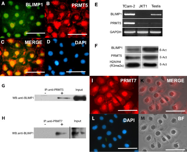
Background: Most testicular germ cell tumors arise from intratubular germ cell neoplasia unclassified (IGCNU, also referred to as carcinoma in situ), which is thought to originate from a transformed primordial germ cell (PGC)/gonocyte, the fetal germ cell. Analyses of the molecular profile of IGCNU and seminoma show similarities to the expression profile of fetal germ cells/gonocytes. In murine PGCs, expression and interaction of Blimp1 and Prmt5 results in arginine 3 dimethylation of histone H2A and H4. This imposes epigenetic modifications leading to transcriptional repression in mouse PGCs enabling them to escape the somatic differentiation program during migration, while expressing markers of pluripotency.

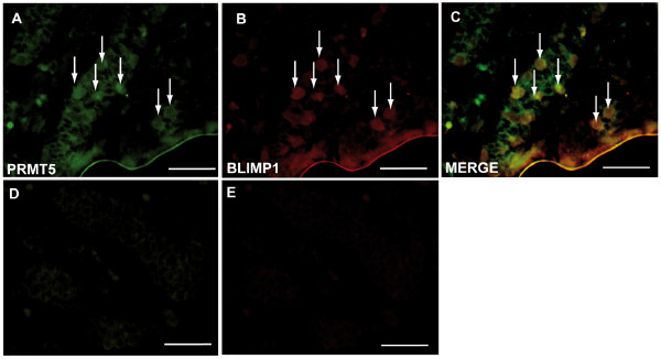
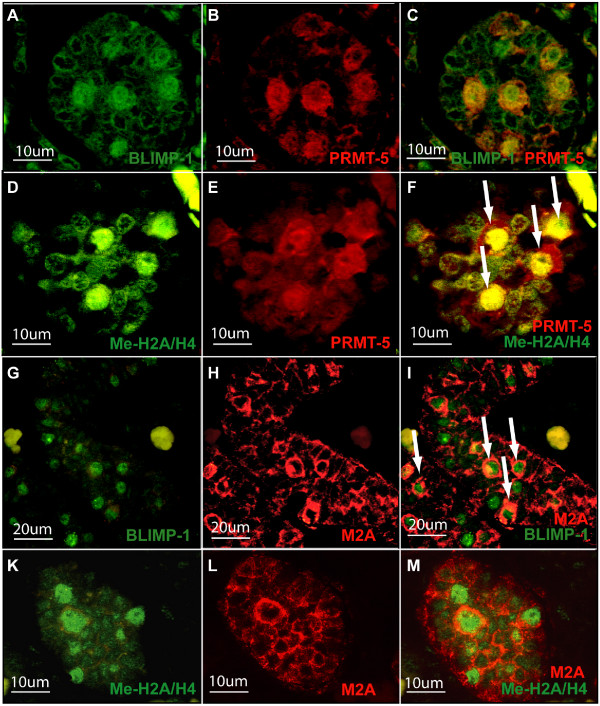
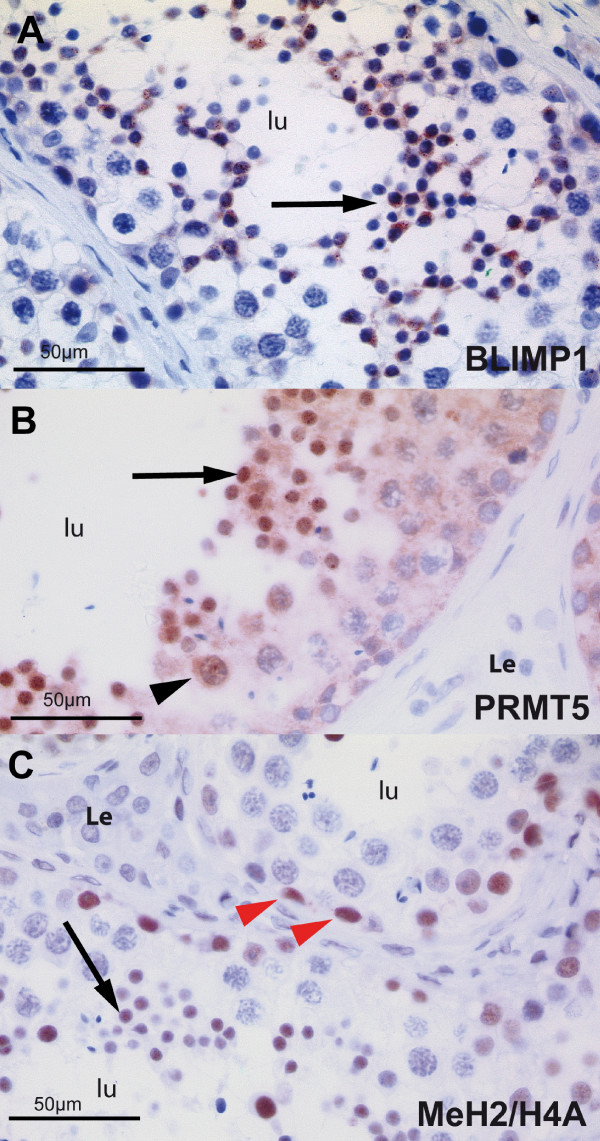
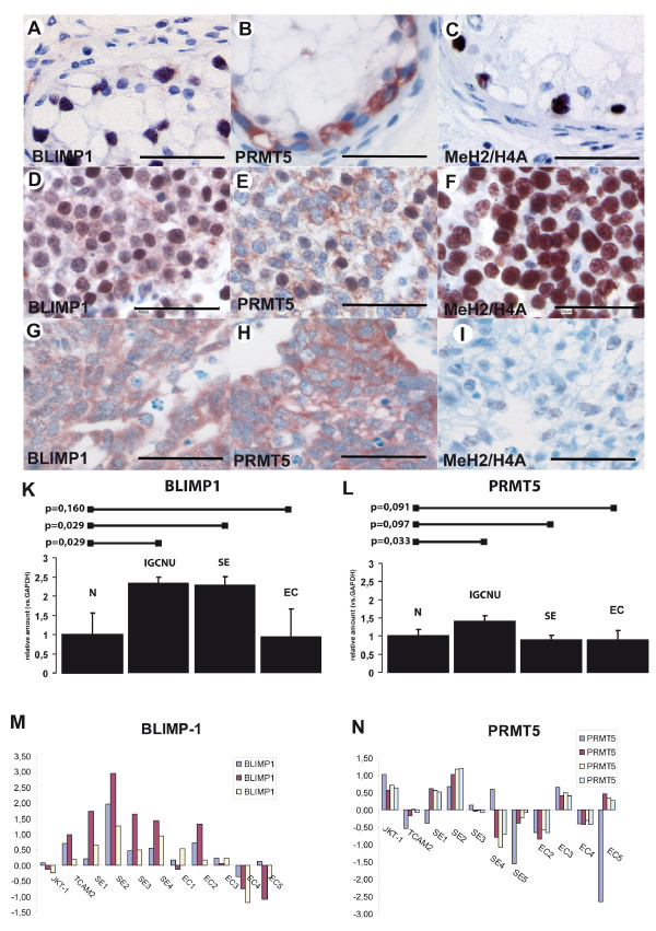
Results: In the present study, we show that BLIMP1 and PRMT5 were expressed and arginine dimethylation of histones H2A and H4 was detected in human male gonocytes at weeks 12-19 of gestation, indicating a role of this mechanism in human fetal germ cell development as well. Moreover, BLIMP1/PRMT5 and histone H2A and H4 arginine 3 dimethylation was present in IGCNU and most seminomas, while downregulated in embryonal carcinoma (EC) and other nonseminomatous tumors.

Conclusion: These data reveal similarities in marker expression and histone modification between murine and human PGCs. Moreover, we speculate that the histone H2A and H4 arginine 3 dimethylation might be the mechanism by which IGCNU and seminoma maintain the undifferentiated state while loss of these histone modifications leads to somatic differentiation observed in nonseminomatous tumors.


Figures

Figure 1
Figure 1
Human fetal gonocytes at 12th week of pregnancy. Sections of human fetal gonocytes at 12th week of pregnancy subjected to antibody staining towards BLIMP1 (A), PRMT5 (B) and overlay (C). D and E no primary antibody controls. Arrows indicate exemplary germ cells. Bar = 50 μm.
Figure 2
Figure 2
Human fetal gonocytes at 19th week of pregnancy. Sections of human fetal gonocytes at 19th week of pregnancy subjected to antibody staining towards (A) BLIMP1, (B) PRMT5, (C) Merge of BLIMP1 and PRMT5, (D) PRMT5, (E) methylated H2A/H4, (F) merge of PRMT5 and methylated H2A/H4 (G) BLIMP1 (H) M2A antigen, (I) merge of BLIMP1 and M2A, (K) methylated H2A/H4, (L) M2A, (M) merge of methylated H2A/H4 and M2A.
Figure 3
Figure 3
Human adult testis. Sections of normal human adult testis stained for BLIMP1 (A), PRMT5 (B) methylated and dimethylated histones H2A/H4 (C). (A) A seminiferous tubule is shown with normal spermatogenesis. Spermatogonia, spermatocytes, and Sertoli cells are devoid of the staining, while nuclear and cytoplasmatic staining occurs in round spermatids (large arrow). (B) Staining with PRMT5 antibody shows low expression of PRMT5 in the nuclei of spermatocytes (arrowhead), and strong nuclear staining in round spermatids (large arrow). (C) Positive staining with Me H2A/H4 occurs in spermatogonia (red arrowheads) and round spermatids (arrow), but not in spermatocytes. lu lumen of the seminiferous tubule; Le Leydig Cells.
Figure 4
Figure 4
Human germ cell tumors. Sections of neoplastic germ cells of IGCNU (A-C), seminoma (D-F), embryonal carcinoma (G-I) stained for BLIMP1 (A, D, G), PRMT5 (B, E, and H) and methylated histones H2A/H4 (C, F, I). In Figure A-C tubules with IGCNU are shown with consistent nuclear expression of BLIMP1 and Me H2A/H4 in neoplastic germ cells (A, BLIMP1; C, Me H2A/H4). PRMT5 is expressed in the cytoplasm of neoplastic germ cells (B). Notice that no expression is present in Sertoli cells. In Figures D-F expression in seminomas is presented. Notice the variation of the expression of BLIMP1, being low or moderate in the majority of the cells (D). PRMT5 is expressed in the cytoplasm of most seminoma cells, but some neoplastic cells also show nuclear staining (E). Figure F shows a strong nuclear staining of MeH2A/H4 in most seminoma cells. Size bar is 50 μm. Quantification of the relative expression of BLIMP1 (K) and PRMT5 (L) normalized to β-Actin and compared to normal testicular tissue. Bars above the graph indicate p-values. (M, N) Expression values for BLIMP1 (M) and PRMT5 (N) from independent Affymetrix expression analyses (as referred in 23). Data are plotted as Log2 (y-axis) after normalization. Abbreviations: Normal testicular tissue (N), IGCNU, seminoma (SE), embryonal carcinoma (EC).
Figure 5
Figure 5
Analysis of TCam-2 seminoma cell line. (A-B) Immunohistochemistry using the antibodies indicated. (C) Merge of (A) and (B). (D) Counterstaining with DAPI to detect nuclei. (E) RT-PCR cell lines TCam2 and JKT1 as well as Testis detecting expression of the indicated genes. (F) Western Blot of protein lysate from TCam2 cells detecting the proteins indicated. (G) Co-IP experiment using antibody to PRMT5 for IP and antibody to BLIMP-1 to detect potential interaction. – no Antibodyl; + IP using PRMT5 Antibody; Input Control. (H) Co-IP experiment using antibody to PRMT7 for IP and antibody to BLIMP-1 to detect potential interaction. – no Antibodyl; + IP using PRMT5 Antibody; Input Control. (I-M) Immunohistochemistry using the PRMT7 antibody (I), (K) Merge of (I) and (M), (L) Counterstaining with DAPI to detect nuclei, (M) brightfield image. Scale Bar indicates 25 μm.

Similar articles

Cited by

References

    1. Lee F, Hamid R, Arya M, Patel HR. Testicular cancer: current update and controversies. Hosp Med. 2002;63:615–20. - PubMed
    1. McGlynn KA, Devesa SS, Graubard BI, Castle PE. Increasing incidence of testicular germ cell tumors among black men in the United States. J Clin Oncol. 2005;23:5757–61. doi: 10.1200/JCO.2005.08.227. - DOI - PubMed
    1. Oosterhuis JW, Looijenga LH. Testicular germ-cell tumours in a broader perspective. Nat Rev Cancer. 2005;5:210–22. doi: 10.1038/nrc1568. - DOI - PubMed
    1. Skakkebaek NE, Berthelsen JG, Giwercman A, Muller J. Carcinoma-in-situ of the testis: possible origin from gonocytes and precursor of all types of germ cell tumours except spermatocytoma. Int J Androl. 1987;10:19–28. doi: 10.1111/j.1365-2605.1987.tb00161.x. - DOI - PubMed
    1. Honecker F, Stoop H, de Krijger RR, Chris Lau YF, Bokemeyer C, Looijenga LH. Pathobiological implications of the expression of markers of testicular carcinoma in situ by fetal germ cells. J Pathol. 2004;203:849–57. doi: 10.1002/path.1587. - DOI - PubMed

Publication types

MeSH terms

LinkOut - more resources