. 2008 Dec 10;582(29):4039-46.
doi: 10.1016/j.febslet.2008.10.043. Epub 2008 Nov 4.

Acute internalization of gap junctions in vascular endothelial cells in response to inflammatory mediator-induced G-protein coupled receptor activation

Affiliations

Acute internalization of gap junctions in vascular endothelial cells in response to inflammatory mediator-induced G-protein coupled receptor activation

Susan M Baker et al. FEBS Lett. .

Abstract

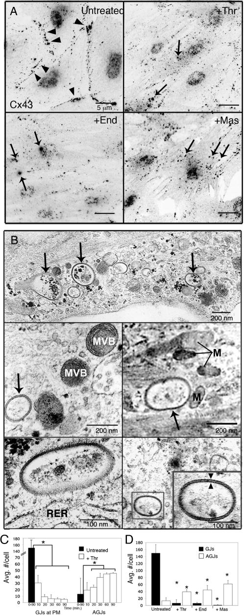
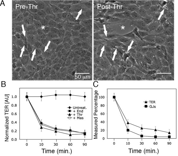
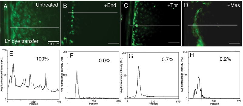
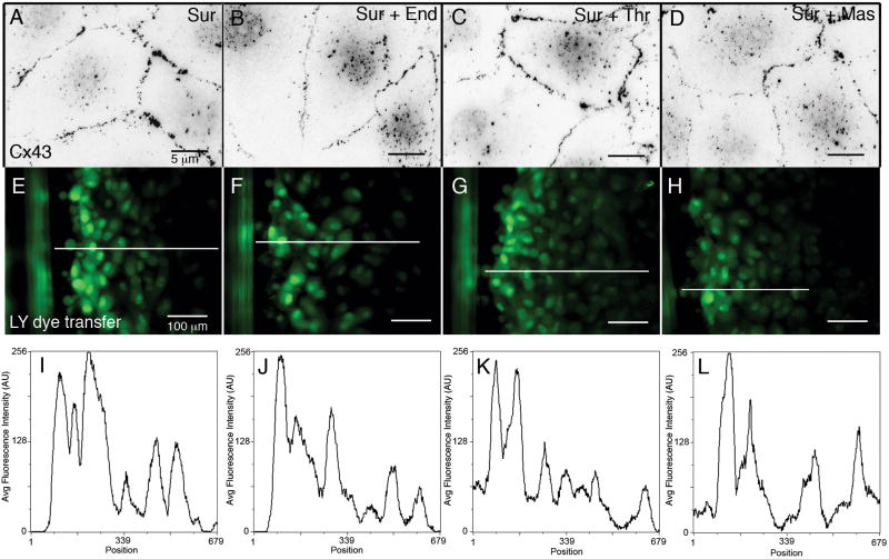
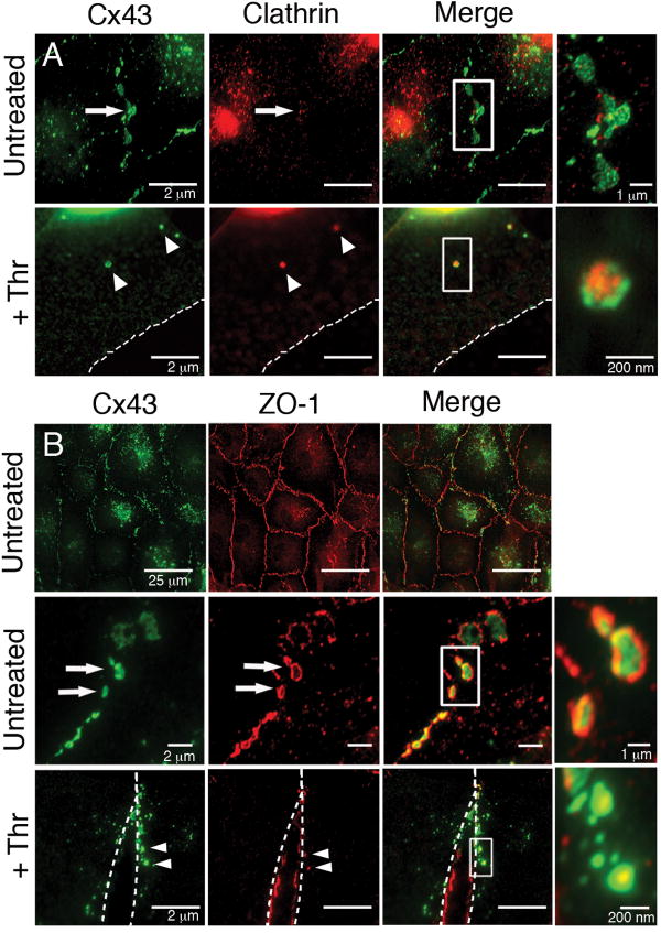
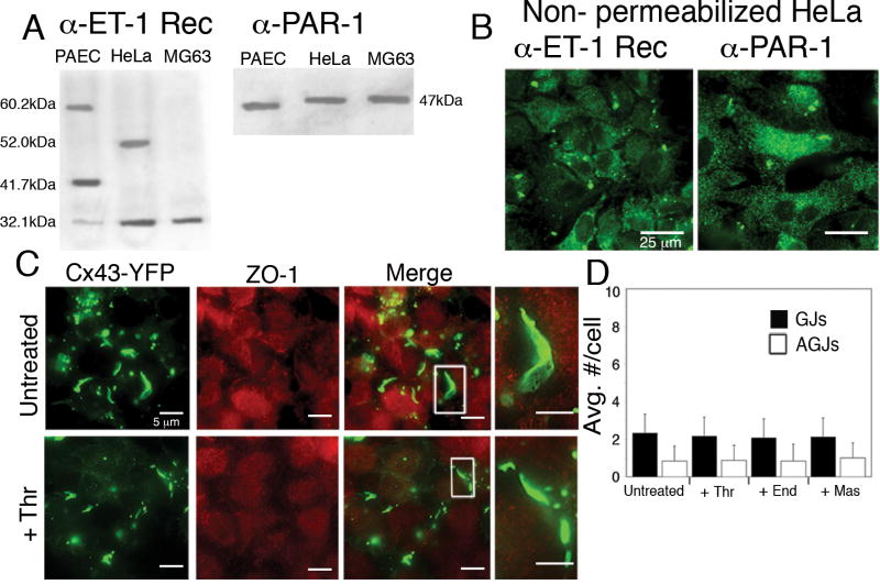
During the inflammatory response, activation of G-protein coupled receptors (GPCRs) by inflammatory mediators rapidly leads to inhibition of gap junction intercellular communication (GJIC); however, the steps that lead to this inhibition are not known. Combining high-resolution fluorescence microscopy and functional assays, we found that activation of the GPCRs PAR-1 and ET(A/B) by their natural inflammatory mediator agonists, thrombin and endothelin-1, resulted in rapid and acute internalization of gap junctions (GJs) that coincided with the inhibition of GJIC followed by increased vascular permeability. The endocytosis protein clathrin and the scaffold protein ZO-1 appeared to be involved in GJ internalization, and ZO-1 was partially displaced from GJs during the internalization process. These findings demonstrate that GJ internalization is an efficient mechanism for modulating GJIC in inflammatory response.


Figures

Figure 1
Figure 1
Effects of inflammatory mediator activation of GPCRs on GJ localization. Primary porcine PAECs were treated with thrombin (10U/mL), endothelin-1 (50nM) or mastoparan (240nM) for 20 min. (A) Immunofluorescence images (black and white inverted for better visibility) of untreated monolayers showed numerous punctate Cx43-based GJs at cell peripheries (Untreated, arrowheads). Activation of GPCRs with inflammatory mediators, thrombin (+Thr), endothelin-1 (+End) or the wasp toxin, mastoparan (+Mas), resulted in loss of GJ signal at the PM and the appearance of numerous intracellular AGJs (arrows) (B) Ultrastructural analysis of AGJs in thrombin-treated PAECs. AGJs are visible near cell borders (upper panel) and deeper in the cytoplasm (middle and lower panels). AGJs exhibited typical double-membrane morphology (lower right panel, inset, arrowheads). (C) Significant reduction of GJs at the PM was accompanied by a significant increase in AGJs in thrombin-treated PAECs over time (n=45 cells), and (D) after 20-minute incubations with thrombin, endothelin-1 and mastoparan (n=15 cells). Data shown are mean ± SEM; p<0.05 (asterisks). MVB=multivesicular body; M=mitochondria; RER=rough endoplasmic reticulum
Figure 2
Figure 2
Vascular endothelial cell permeability increases in response to inflammatory mediators. (A) Phase contrast images of a single field of view of a PAEC monolayer pre- and post-thrombin (10U/mL) treatment. Note the development of smaller separations at arrows and a larger “gap” (asterisk) within 30 minutes, as cells physically separated from one another. (B) TER of PAECs was determined over time before and after addition of endothelin-1 (50nM), thrombin (10U/mL) or mastoparan (240nM) Resistance measurements of untreated cells were used to establish a baseline for normalization, and (C) average number of GJs at the PM (n=15 cells) and average TER measurements (n=9) in treated cells were compared to untreated cells and expressed as a percentage. Values shown are mean ± SEM.
Figure 3
Figure 3
Inhibition of GJIC coincides with GJ internalization in response to GPCR activation by inflammatory mediators. (A-D) GJIC was assessed by scrape loading dye transfer in untreated PAEC monolayers (A); and following 30-min. incubations with 50nM endothelin-1 (B), 10U/mL thrombin (C), or 240nM mastoparan (D). (E-H) Scans of fluorescence intensity measured along lines indicated in A-D. The area under the curve for untreated cells was set as 100% dye transfer, and the areas of the curves for treated cells were expressed as a percentage of untreated control.
Figure 4
Figure 4
Effects of blocking GPCR activation in the presence of inflammatory mediators. (A-D) Immunofluorescence images of Cx43-based GJs showed that neither GJ internalization nor cell separation occurred in PAECs treated with 240nM suramin alone (A), or with suramin plus 50nM endothelin-1 (B), 10U/mL thrombin (C), or 240nM mastoparan (D) for 30 min. (E-H) GJIC was assessed by scrape loading dye transfer in PAECs treated under the same conditions as in A-D. (I-L) Linescans of average fluorescence intensity measured along the lines indicated in E-H.
Figure 5
Figure 5
Colocalization of Cx43 with the endocytic coat protein clathrin and the scaffold protein ZO-1. (A) Immunofluorescence colocalization showed a patchy distribution of clathrin (red) at Cx43-based GJ plaques (green) in PAECs (Untreated, arrows; see merged inset) and colocalized with internalized Cx43 AGJs following 20-min. incubations with 10U/mL thrombin (+Thr, arrowheads; see merged inset). (B) Immunofluorescence colocalization showed robust colocalization of ZO-1 (red) at virtually all Cx43-based GJ plaques (green) in untreated PAECs (Untreated, upper panel) particularly at plaque peripheries (Untreated, lower panel, arrows; see merged inset). In PAECs treated 20 min. with thrombin, ZO-1 remained localized at the PM of separating cells (+Thr, dashed outlines), and appeared to be localized preferentially at the center of Cx43 AGJs (+Thr, arrowheads; see merged inset).
Figure 6
Figure 6
ZO-1-Cx43 interaction is required for GJ internalization in response to inflammatory mediators. (A, B) Western blot (A) and immunofluorescence (B) of HeLa-22 cells stably expressing Cx43-YFP confirmed the expression of both endothelin-1 receptors (α-ET-1 Rec) and thrombin receptors (α-PAR-1). (C) Immunofluorescence colocalization of ZO-1 (red) with Cx43-YFP (green) showed no colocalization in untreated HeLa22 cells (Untreated; see merged inset) or in HeLa22 cells treated 20 min. with thrombin (+Thr; see merged inset). (D) No increase in GJ internalization in response to thrombin, endothelin-1 or mastoparan treatment for 20 min. was observed in the absence of ZO-1 binding in HeLa22 cells (n=15 cells). Values shown are mean ± SEM.

References

    1. Mehta PP, Bertram JS, Loewenstein WR. Growth inhibition of transformed cells correlates with their junctional communication with normal cells. Cell. 1986;44:187–96. - PubMed
    1. Loewenstein WR, Rose B. The cell-cell channel in the control of growth. Semin Cell Biol. 1992;3:59–79. - PubMed
    1. Guthrie SC, Gilula NB. Gap junctional communication and development. Trends Neurosci. 1989;12:12–6. - PubMed
    1. Falk M, Baker SM, Segretain D, Buckheit RW., III Gap junction turnover is achieved by the release of small endocytic double-membrane vesicles that coincides with channel accrual. PNAS. 2008 in revision. - PMC - PubMed
    1. Piehl M, Lehmann C, Gumpert A, Denizot JP, Segretain D, Falk MM. Internalization of large double-membrane intercellular vesicles by a clathrin-dependent endocytic process. Mol Biol Cell. 2007;18:337–47. - PMC - PubMed

Publication types

MeSH terms