. 2008 Nov;15(11):1360-75.
doi: 10.1016/j.acra.2008.07.007.

Multivariate analysis of structural and diffusion imaging in traumatic brain injury

Affiliations

Multivariate analysis of structural and diffusion imaging in traumatic brain injury

Brian Avants et al. Acad Radiol. 2008 Nov.

Abstract

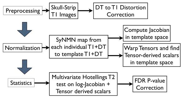
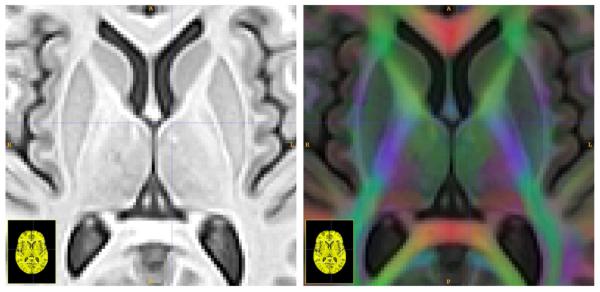
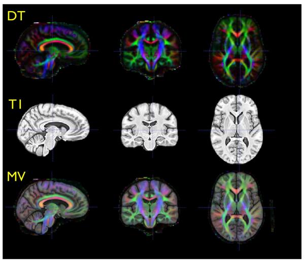
Rationale and objectives: Diffusion tensor (DT) and T1 structural magnetic resonance images provide unique and complementary tools for quantifying the living brain. We leverage both modalities in a diffeomorphic normalization method that unifies analysis of clinical datasets in a consistent and inherently multivariate (MV) statistical framework. We use this technique to study MV effects of traumatic brain injury (TBI).

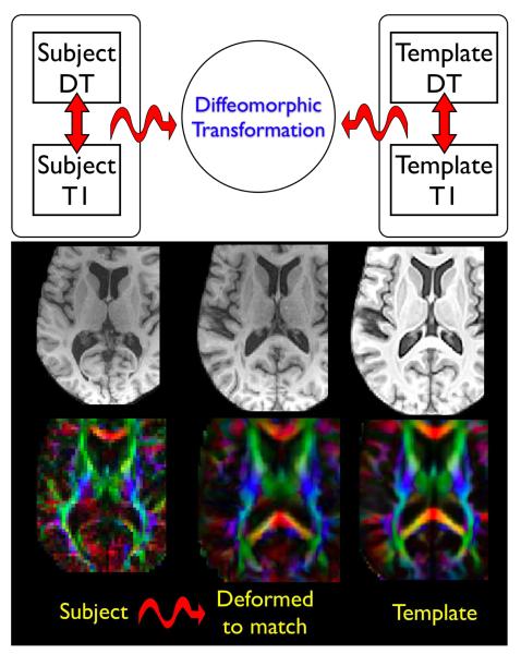
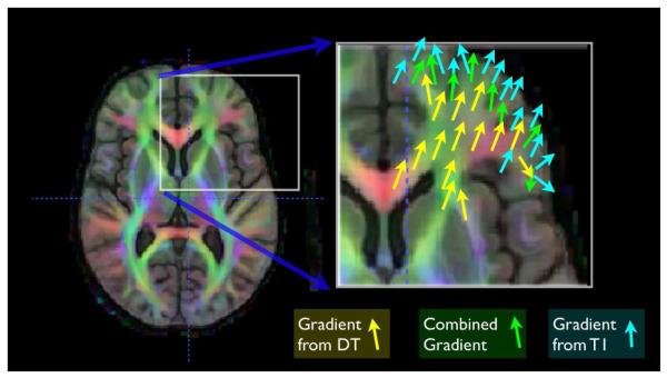
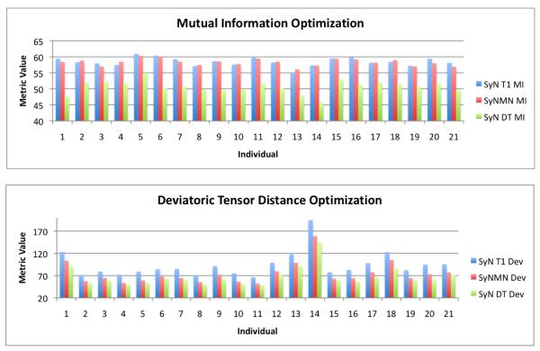
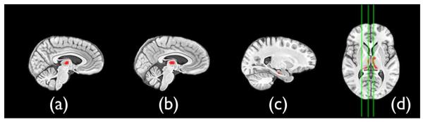
Materials and methods: We contrast T1 and DT image-based measurements in the thalamus and hippocampus of 12 TBI survivors and nine matched controls normalized to a combined DT and T1 template space. The normalization method uses maps that are topology-preserving and unbiased. Normalization is based on the full tensor of information at each voxel and, simultaneously, the similarity between high-resolution features derived from T1 data. The technique is termed symmetric normalization for MV neuroanatomy (SyNMN). Voxel-wise MV statistics on the local volume and mean diffusion are assessed with Hotelling's T(2) test with correction for multiple comparisons.

Results: TBI significantly (false discovery rate P < .05) reduces volume and increases mean diffusion at coincident locations in the mediodorsal thalamus and anterior hippocampus.

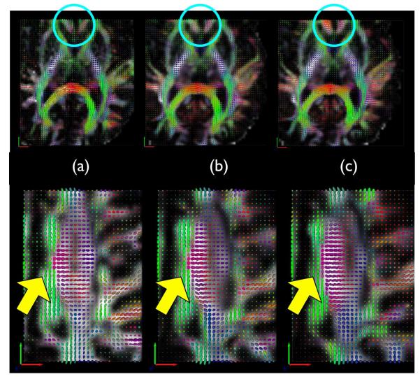
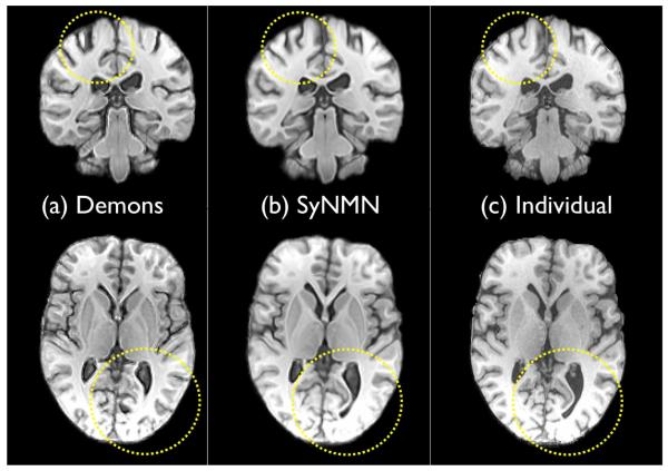
Conclusions: SyNMN reveals evidence that TBI compromises the limbic system. This TBI morphometry study and an additional performance evaluation contrasting SyNMN with other methods suggest that the DT component may aid normalization quality.


Figures

Fig. 1
Fig. 1
An overview of the steps required in our multivariate processing for population analysis of tensor and scalar components. The Jacobian measures local volume of neuroanatomy, while tensor-derived scalars relate to the integrity of cellular structure. Multivariate tests enable relationships between these variables to enhance statistical power for detecting group differences.
Fig. 2
Fig. 2
Here we see one slice from the template T1 (left) and T1 plus DT (right) after intra-modality distortion correction. The RGB channel intensity is weighted by the FA and indicates principal fiber direction going left-to-right (red) going bottom-to-top (green) and going in-and-out of the page (blue) which are superimposed on the T1 image in the right panel.
Fig. 3
Fig. 3
Triplanar views of the DT component of the template (top), the T1 component of the template (center) and the combined MV template (bottom). Inter– modality distortion correction, with a small deformation model, is necessary to bring these images into alignment.
Fig. 4
Fig. 4
Two integrated MV datasets of individual anatomical structure and the mapping between the MV datasets via transformation, ϕ. Our methods are able to leverage both modalities to guide the computation of ϕ with maximal subject information. Curved arrows indicate intersubject transformations while straight arrows indicate intrasubject transformations. The TBI subject, at left, exhibits ventricular enlargement visible in both the DT and T1 modality. Note that the SyNMN technique is able to deform both modalities into the configuration shown in the center column, closely resembling the template in the right column. This map factors out diffeomorphic shape differences between the individual brain volumes.
Fig. 5
Fig. 5
The optimization of the DT and T1 combined similarity term leads to a combined gradient term. Within white matter, the majority of the gradient contribution comes from the DT component, while elsewhere the gradient is either from the T1 or a smooth combination of T1 and DT, at white matter and gray matter interfaces.
Fig. 6
Fig. 6
Column (a) shows a target DT image where as (b) shows a SyNMN mapping from an individual DT to the target DT. Column (c) shows the SyN result, which ignores the DT component. Better alignment of tensors with SyNMN are visible within the caudate at top and within the corticospinal tract along the bottom row. The lack of quality tensor alignment in the SyN result is not due to low quality normalization of T1 images, but rather due to the fact that the data visualized here is invisible in the T1 modality.
Fig. 7
Fig. 7
The bivariate performance, in our TBI dataset, of SyN-T1, SyN-DT and SyNMN are shown in the graphs above for mutual information and the deviatoric tensor distance. SyNMN provides a balanced optimization of both terms, across the dataset. For some cases, one may see that SyNMN outperforms SyN in the mutual information term.
Fig. 8
Fig. 8
Two slices from the Demons mapping of template to individual are shown in (a); the SyNMN mapping of template to individual is in (b). The individual is in (c). Note that the Demons mapping is of overall high quality. However, the fine details (some outlined in yellow) are not as accurately mapped as in SyN and SyNMN. Detailed matching – and the ability to capture large deformation – is where the advantage of diffeomorphisms arise, in contrast to “elastic” types of methods. However, we cannot claim to have an optimal implementation of the Demons algorithm.
Fig. 9
Fig. 9
T2 results after FDR correction at p < 0.05 indicate that the left and right mediodorsal thalamus and the hippocampus are affected by TBI. Sagittal slices (a) (b) and (c) are highlighted in the axial slice and indicate right mediodorsal thalamus, left mediodorsal thalamus and the left anterior hippocampus, respectively. The MV test, here, indicates that the mean diffusion is larger and the log-Jacobian smaller in the TBI subjects relative to controls. Similar results were gained with SyN. However, the hippocampus cluster did not survive FDR correction. Images are displayed in radiologic convention.

Similar articles

Cited by

References

    1. Furlow Bryant. Diagnostic imaging of traumatic brain injury. Radiol Technol. 2006;78:145–56. quiz 157-9. - PubMed
    1. Hoge Charles W, McGurk Dennis, Thomas Jeffrey L, Cox Anthony L, Engel Charles C, Castro Carl A. Mild traumatic brain injury in U.S. Soldiers returning from Iraq. N Engl J Med. 2008;358:453–463. - PubMed
    1. Kraus Marilyn F, Susmaras Teresa, Caughlin Benjamin P, Walker Corey J, Sweeney John A, Little Deborah M. White matter integrity and cognition in chronic traumatic brain injury: a diffusion tensor imaging study. Brain. 2007;130:2508–2519. - PubMed
    1. Mendez Cecilia V, Hurley Robin A, Lassonde Maryse, Zhang Liying, Taber Katherine H. Mild traumatic brain injury: neuroimaging of sports-related concussion. J Neuropsychiatry Clin Neurosci. 2005;17:297–303. - PubMed
    1. Chen Jen-Kai, Johnston Karen M, Petrides Michael, Ptito Alain. Neural substrates of symptoms of depression following concussion in male athletes with persisting postconcussion symptoms. Arch Gen Psychiatry. 2008;65:81–89. - PubMed

Publication types