Comment
. 2008 Nov 6;60(3):398-9.
doi: 10.1016/j.neuron.2008.10.030.

N-Cadherin and integrins: two receptor systems that mediate neuronal process outgrowth on astrocyte surfaces

Affiliations
Comment

N-Cadherin and integrins: two receptor systems that mediate neuronal process outgrowth on astrocyte surfaces

Louis F Reichardt. Neuron. .

Abstract

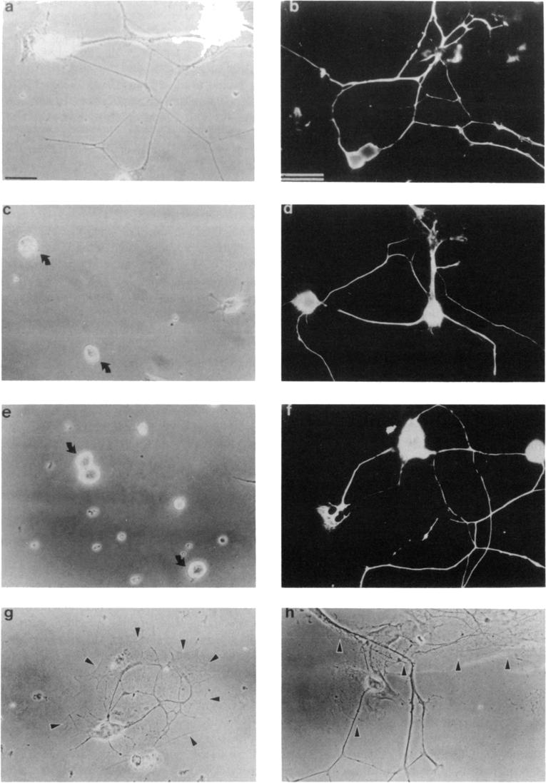
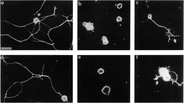
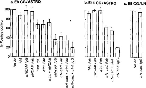
Receptor-mediated interactions between neurons and astroglia are likely to play a crucial role in the growth and guidance of CNS axons. Using antibodies to neuronal cell surface proteins, we identified two receptor systems mediating neurite outgrowth on cultured astrocytes. N-cadherin, a Ca2+-dependent cell adhesion molecule, functions prominently in the outgrowth of neurites on astrocytes by E8 and E14 chick ciliary ganglion (CC) neurons. β1-class integrin ECM receptor heterodimers function less prominently in E8 and not at all in E14 neurite outgrowth on astrocytes. The lack of effect of integrin β1 antibodies on E14 neurite outgrowth reflects an apparent loss of integrin function, as assayed by E14 neuronal attachment and process outgrowth on laminin. N-CAM appeared not to be required for neurite outgrowth by either E8 or E14 neurons. Since N-cadherin and integrin β1 antibodies together virtually eliminated E8 CG neurite outgrowth on cultured astrocytes, these two neuronal receptors are probably important in regulating axon growth on astroglia in vivo.


Figures

Figure 1
Figure 1. E8 and E14 CG Neuronal Process Outgrowth on Primary Cortical Astrocytes and on Substrates Coated with Astrocyte CM
Neurons were visualized either by phase contrast or, when grown on astrocyte monolayers, by immunofluorescent staining with the A2B5 monoclonal antibody. (a) E8 CG neurons grown 16 hr on an astrocyte CM substrate. (b) E8 CG neurons grown 16 hr on a monolayer of astrocytes. (c) E8 CG neurons grown 16 hr on an astrocyte CM substrate in the presence of 100 μg/ml anti-integrin β1 (JG22) IgG. Note process outgrowth is inhibited. (d) E8 CG neurons grown 16 hr on astrocyte surfaces in the presence of 1 μg/ml anti-integrin β1 (JG22) Fab. Note process outgrowth is not prevented as it is on astrocyte CM (e) E14 CG neurons grown 16 hr on astrocyte CM. Arrows mark E14 neurons that do not respond to astrocyte CM (f) E14 CG neurons grown 16 hr on astrocyte surfaces. Note E14 CG neurons extend long neurites on astrocyte surfaces. (g) E14 CG neurons cultured 16 hr on small astrocyte islands centered on laminin-coated, glass coverslips. Note E14 CG neurites do not extend off of the astrocyte monolayer. (h) In contrast to E14 neurites, E8 neurites often grow off the astrocyte islands onto the surrounding laminin substrates. Arrowheads in (g) and (h) mark the boundary between the astrocyte monolayer and the surrounding laminin substrate. Bars, 10 μm. Bar in (a) also applies to (c) and (e); bar in (b) also applies to (d) and (f-h).
Figure 2
Figure 2. E8 and E14 CG Neuronal Process Outgrowth on Astrocyte Surface Is Strongly Inhibited by Anti-N-Cadherin but Not Anti-N-CAM
Neurons were grown 16 hr on astrocyte surfaces in the presence of polyclonal anti-N-CAM IgG (a and d; 1 mg/ml), anti-N-cadherin IgG (b and c; 1 mg/ml), or anti-N-cadherin Fab’ (e and f; 1 mg/ml). The majority of E8 (a-c) and E14 (d-f) CG neurons were prevented from initiating neurites in the presence of N-cadherin antibodies. However, short neurites with enlarged growth cones were sometimes seen (arrows in c and f). Process outgrowth in the presence of anti-N-CAM appears to be unaffected (a and d). Neurons were visualized by immunofluorescent staining with the A2B5 monoclonal antibody and were readily distinguished from A2B5-positive astrocytes using morphological criteria. Bar, 10 μm.
Figure 3
Figure 3. Distribution of the Lengths of CG Neurites Extended on Astrocyte Surfaces in the Presence of Antibodies to N-Cadherin. N-CAM, and Integrin β1
E8 (a and b) or E14 (c) CG neurons were cultured 16−20 hr on astrocyte monolayers in the presence of different antibodies, and camera lucida tracings of fixed and stained neurites were measured. The percentage of neurons with neurites greater than a certain length (vertical axis) is expressed as a function of neurite length (horizontal axis). Each graph consists of determinations made on individual cultures from the same experiment. (a) Distribution of neurite lengths of E8 CG neurons grown on astrocytes in the presence of 1A6 IgG (∼500 μg/ml; Δ), anti-integrin β1 (CSAT) IgG (100 μg/ml; ▲), anti-N-cadherin IgG (1 mg/ml; ◆), or anti-integrin β1 (CSAT) IgG plus anti-N-cadherin IgG (100 μg/ml and 1 mg/ml, respectively; ◇). In this experiment, 1A6 IgG served as a control antibody since it has no significant effect on neurite outgrowth on astrocyte surfaces (see Figures 4a and 4b). (b) Distribution of neurite lengths of E8 CG neurons grown on astrocytes in the absence of antibodies (△) or in the presence of anti-N-CAM Fab’ (1 mg/ml; ■), anti-integrin β1 (JG22) Fab’ (1 mg/ml; ▲), anti-integrin β1 (JG22) Fab plus anti-N-CAM Fab’ (1 mg/ml each; □), anti-N-cadherin Fab’ (1 mg/ml; ◆), or anti-integrin β1 (JG22) Fab plus anti-N-cadherin Fab’ (1 mg/ml each; ◇). (c) Distribution of neurite lengths of E14 CG neurons grown on astrocytes in the absence of antibody (Δ) or in the presence of anti-N-CAM Fab’ (1 mg/ml; ■), anti-integrin β1 (JG22) Fab (1 mg/ml; ▲), anti-N-cadherin Fab’ (1 mg/ml; ◆), anti-integrin Fab’ plus anti-integrin β1 (JG22) Fab (1 mg/ml each; ◇), or anti-N-cadherin IgG (1 mg/ml; □). Symbol Summary: △, control; ▲, anti-integrin β1; ◆, anti-N-cadherin; ◇, anti-integrin β1 plus anti-N-cadherin; ■, anti-N-CAM; □, anti-N-CAM plus anti-integrin β1 in (b), anti-N-cadherin IgG in (c).
Figure 4
Figure 4. Average Total Length of Neurites Extended by E8 or E14 CG Neurons on Astrocytes or on Laminin in the Presence of Different Antibodies
Values are expressed as percentage of the neurite length in the absence of added antibodies (positive control). In (a) error bars represent the standard error of determinations made on 30−35 neurons per condition. In (b), error bars represent the range of average neurite lengths from two different experiments (total 60−70 neurons per condition). (a) E8 CG neurons were grown 16−20 hr on astrocytes in the absence of antibodies or in the presence of 1A6 Fab (1 mg/ml), anti-N-CAM IgG (1 mg/ml), anti-N-CAM Fab’ (1 mg/ml), anti-integrin β1 (CSAT) IgG (100 μg/ml), anti-integrin β1 (JG22) Fab (1 mg/ml), anti-integrin β1 (JG22) Fab plus anti-N-CAM Fab’ (1 mg/ml each), anti-N-cadherin IgG (1 mg/ml), anti-N-cadherin Fab’ (1 mg/ml), anti-N-cadherin Fab’ plus anti-integrin β1 (JG22) Fab (1 mg/ml each), or anti-N-cadherin IgG plus anti-integrin β1 (CSAT) IgG (1 mg/ml and 100 μg/ml, respectively). (b) E14 CG neurons were grown 16−20 hr on astrocytes in the presence of 1A6 Fab (1 mg/ml), anti-N-CAM Fab’ (1 mg/ml), anti-integrin β1 (JG22) Fab (1 mg/ml), anti-N-cadherin Fab’ (1 mg/ml), anti-N-cadherin Fab’ plus anti-integrin β1 (JG22) Fab (1 mg/ml each), or anti-N-cadherin IgG (1 mg/ml). (c) E8 CG neurons were grown 16 hr on laminin substrates in the absence of added antibodies or in the presence of 1 mg/ml anti-N-cadherin IgG.

Comment on

References

    1. Akers RM, Mosher DF, Lilien JE. Promotion of retinal neurite outgrowth by substratum-bound fibronectin. Dev. Biol. 1981;86:179–188. - PubMed
    1. Antonicek H, Persohn E, Schachner M. Biochemical and functional characterization of a novel neuron-glia adhesion molecule that is involved in neuronal migration. J. Cell Biol. 1987;104:1587–1595. - PMC - PubMed
    1. Asch ASL, Leung LK, Shapiro J, Nachman RL. Human brain glial cells synthesize thrombospondin. Proc. Natl. Acad. Sci. USA. 1986;83:2904–2908. - PMC - PubMed
    1. Assouline JG, Bosch P, Lim R, Kim IS, Jensen R, Pantagis NJ. Rat astrocytes and Schwann cells in culture synthesize nerve growth factor-like neurite-promoting factors. Brain Res. 1987;428:103–118. - PubMed
    1. Benfey M, Aguayo AJ. Extensive elongation of axons from rat brain into peripheral nerve grafts. Nature. 1982;296:150–152. - PubMed