

Review
doi: 10.1016/j.neuron.2008.10.020.
Strength through diversity
Affiliations
- PMID: 18995822
- PMCID: PMC4919814
- DOI: 10.1016/j.neuron.2008.10.020
Item in Clipboard
Review
Strength through diversity
Neuron.
.
Abstract
The remarkable versatility of the mammalian brain is made possible by a huge diversity of cellular plasticity mechanisms. These include long-term potentiation and depression at both excitatory and inhibitory synapses, as well as a variety of intrinsic and homeostatic plasticity mechanisms. A fundamental challenge for the field is to assemble our detailed knowledge of these specific mechanisms into a coherent picture of how plasticity within cortical circuits works to tune network properties.
Figures
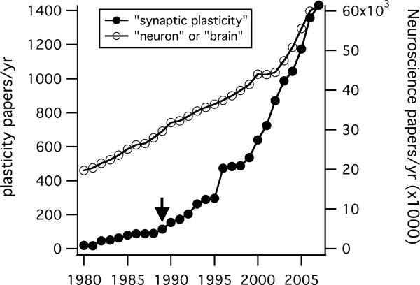
Filled symbols plot the number of papers published per year (left axis) estimated from a Medline search for the terms “synaptic” and “plasticity.” For much of this period (1980–2007) growth outstripped the broader neuroscientific literature estimated from searches for the terms “neuron” or “brain” (right axis). Neural plasticity papers accounted for 1/300 of this larger literature during the 1980s, but account for 1/42 papers in 2007. Arrow marks the year Neuron was founded.
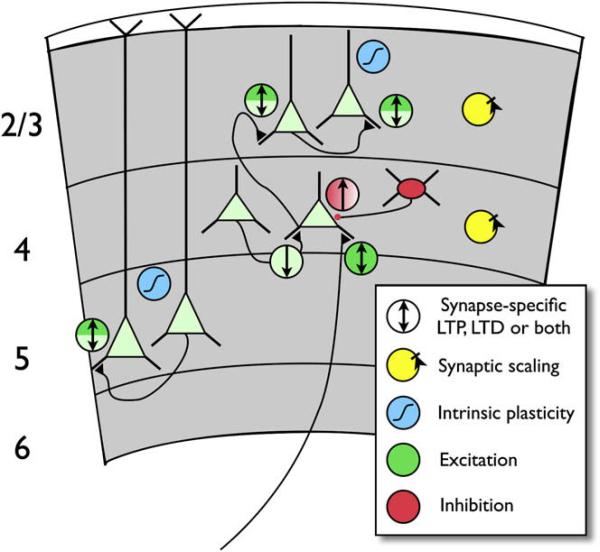
A subset of known synaptic connections between excitatory (pale green) and inhibitory (red) neurons (black triangles are excitatory connections; red dot, inhibitory). Circles with arrows indicate LTP (up arrow), LTD (down arrow), or coexisting LTP and LTD (bidirectional arrow) of excitation (green) or inhibition (red). Light green indicates LTP/D believed to be primarily or exclusively presynaptic, dark green indicates postsynaptic. Both colors indicate evidence for mixed or coexisting pre- and postsynaptic mechanisms. Locus of LTP of inhibition is not known (red gradient). Axon entering layer 4 from below layer 6 indicates thalamocortical input to L4 spiny stellate cell. Excitatory and inhibitory synapses in layers 2/3 and 4 also exhibit bidirectional synaptic scaling (yellow symbols). Intrinsic plasticity (blue symbols) has been demonstrated in L2/3 and L5 pyramidal neurons. Some forms of plasticity shown are developmentally regulated.
References
Publication types
MeSH terms
Grants and funding
LinkOut - more resources
Full Text Sources
Other Literature Sources
