. 2008 Nov 7;32(3):426-38.
doi: 10.1016/j.molcel.2008.10.012.

Hierarchical regulation of WASP/WAVE proteins

Affiliations

Hierarchical regulation of WASP/WAVE proteins

Shae B Padrick et al. Mol Cell. .

Abstract

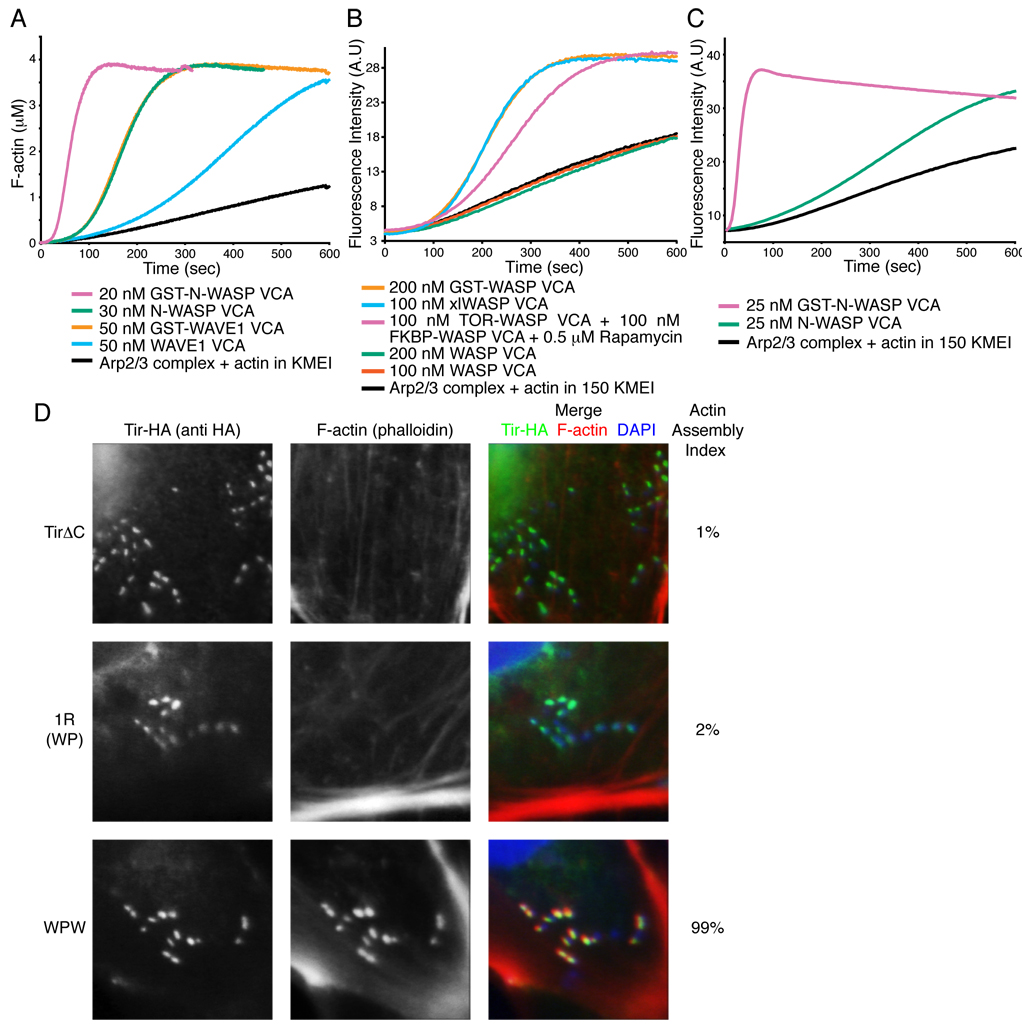
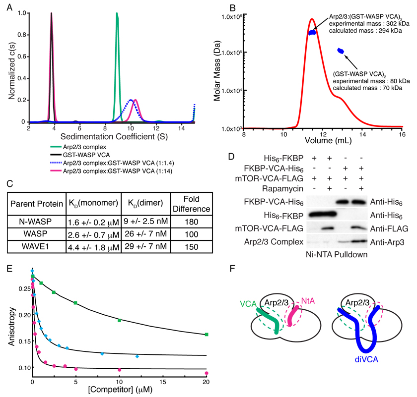
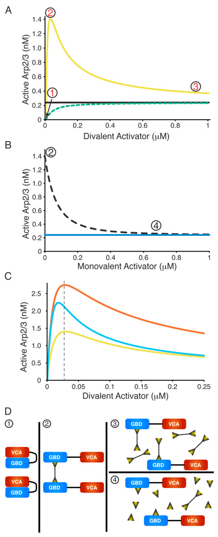
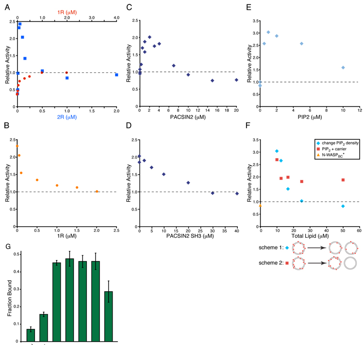
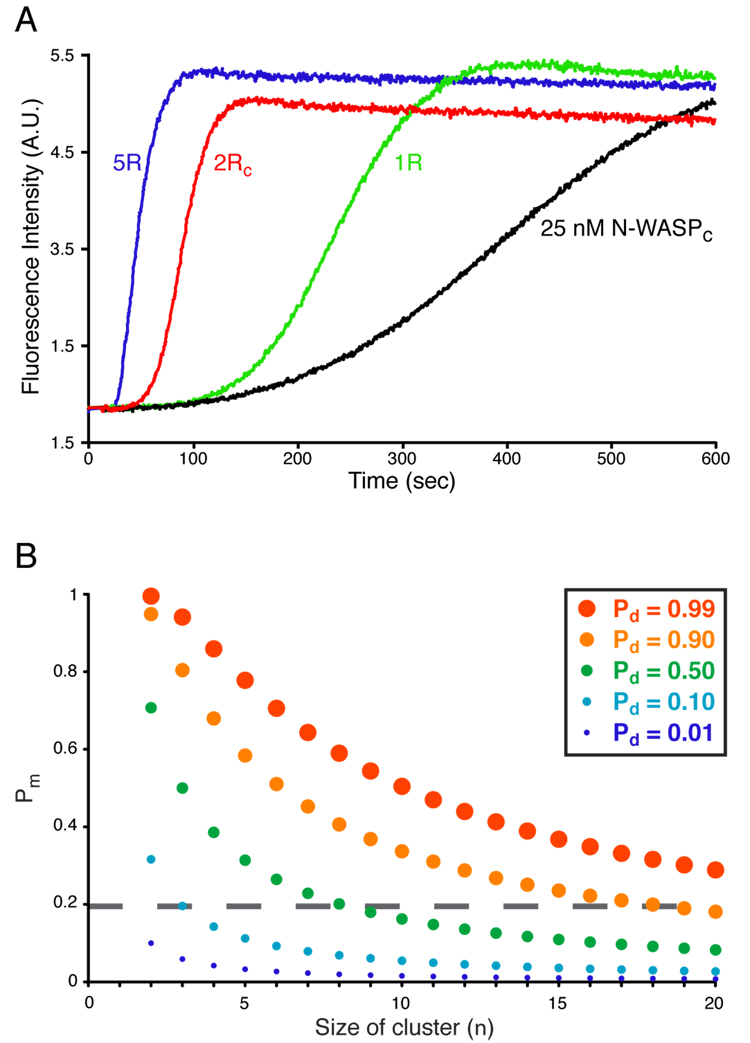
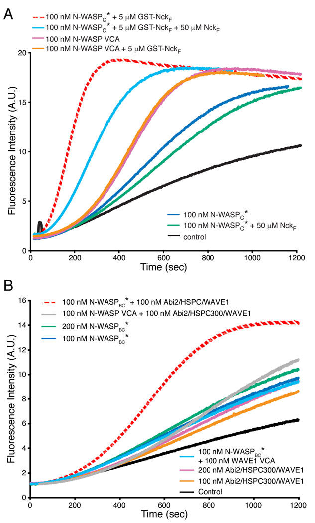
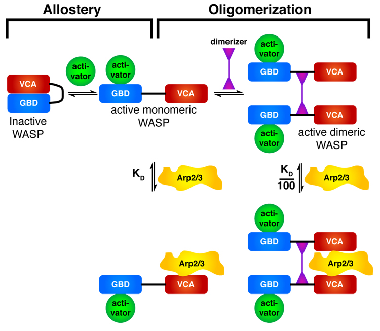
Members of the Wiskott-Aldrich syndrome protein (WASP) family control actin dynamics in eukaryotic cells by stimulating the actin nucleating activity of the Arp2/3 complex. The prevailing paradigm for WASP regulation invokes allosteric relief of autoinhibition by diverse upstream activators. Here we demonstrate an additional level of regulation that is superimposed upon allostery: dimerization increases the affinity of active WASP species for Arp2/3 complex by up to 180-fold, greatly enhancing actin assembly by this system. This finding explains a large and apparently disparate set of observations under a common mechanistic framework. These include WASP activation by the bacterial effector EspFu and a large number of SH3 domain proteins, the effects on WASP of membrane localization/clustering and assembly into large complexes, and cooperativity between different family members. Allostery and dimerization act in hierarchical fashion, enabling WASP/WAVE proteins to integrate different classes of inputs to produce a wide range of cellular actin responses.


Figures

Figure 1
Figure 1. Dimerization Increases VCA Activity In Vitro and in Cells
(A – C) Pyrene fluorescence measured during assembly of actin by Arp2/3 complex (black) plus the indicated components. Without rapamycin, 100 nM mTOR-WASP VCA + 100 nM FKBP-WASP VCA has activity comparable to 200 nM WASP VCA (not shown). (D) Clustered EspFu leads to actin rich pedestal formation, but requires the ability to recruit multiple WASP molecules. Cells expressing HA-tagged TirΔC, TirΔC-1R (WP) or TirΔC-WPW were treated with intimin-expressing E. coli, and stained with anti-HA antibody (green in merged image) and Alexa568-phalloidin (red in merged image). Colocalization is yellow in merged image. The fraction of transfected cells harboring at least five F-actin foci was quantified (Actin Assembly Index). Bacteria were visualized by DAPI staining (blue in merged image). Data represent mean from three independent samples of 30 cells each, standard deviation for all samples is 2%.
Figure 2
Figure 2. VCA Dimers Bind Arp2/3 with 1:1 Stoichiometry and High Affinity
(A) Normalized c(s) distributions from sedimentation velocity ultracentrifugation analyses of GST-WASP VCA (black), Arp2/3 complex (green), and Arp2/3 complex + GST-WASP VCA (0.5 µM + 6.8 µM, magenta; 1.8 µM + 2.6 µM, dashed blue). The black, green, and red distributions were normalized to a maximum c(s) value of 1.0; the blue distribution was normalized to give maximum c(s) value equal to that of the 10.3-S peak of the red distribution. (B) Molar mass distribution of multi-angle light scattering data collected on a 1:3 mixture of Arp2/3 complex and GST-WASP VCA, where excess GST-WASP VCA is resolved using Superdex200. Mass distribution within selected peaks is in blue, chromatogram is shown as red curve (refractive index change). (C) Dissociation constants (KD) for the interaction of Arp2/3 complex with VCA monomers and GST-VCA dimers from N-WASP, WASP and WAVE1. Errors shown are 1σ confidence intervals from fitting. (D) Dimerized VCA increases Arp2/3 complex binding in cells. HEK 293 cells transfected with the indicated constructs were treated with rapamycin or DMSO for 30 minutes and lysed. Complexes were precipitated with NiNTA affinity resin and analyzed by western blotting for the indicated proteins. (E) Competition displacement of cortactin from Arp2/3 complex. 10 nM rNtA was displaced from 200 nM Arp2/3 using the indicated concentrations of NtA (magenta circles), xlWASP VCA (cyan diamonds) or WASP VCA (green squares). Black curves are best fit solutions to the competition binding equilibrium equations (see Supplemental Materials). (F) Cartoon model of (VCA)2 binding to two distinct sites on Arp2/3 complex. The cartoon does not show the exact location of the sites on Arp2/3, but merely the presence of two binding sites, both of which are engaged by di-VCA materials.
Figure 3
Figure 3. Modeling Predicts Unusual Behavior in Dimerizer Titrations
The concentration of active Arp2/3 complex was modeled as a function of dimerizing WASP ligands with different properties. Here, ligand binding is concomitant with allosteric relief of autoinhibition. See Figure S9 for ligands binding to constitutively active WASP ligands. (A) Simulated titrations of monomeric WASP ligand (dashed green, e.g. EspFu 1R) or dimeric WASP ligand (yellow, e.g. EspFu 2R) into WASP plus Arp2/3 complex. Black line indicates activity of VCA. (B) Simulated titration of monomeric WASP ligand (dotted black) into WASP hyperactivated by dimeric WASP ligand (maximum activity in A). Blue line indicates activity of VCA. (C) Simulated titrations of different dimerizing ligands into WASP plus Arp2/3 complex. Titration from (A) is yellow (note change of X- and Y-scales). Sky blue curve models a dimerizer with 3-fold tighter affinity for WASP than in (A), red curve models a dimerizer:(WASP)2 complex with three-fold tighter affinity for Arp2/3 than in (A). Modeling based on 25 nM WASP, 10 nM Arp2/3 complex throughout, and the following affinities in (A) and (B)—ligand:WASP, 30 nM; active monomeric WASP:Arp2/3, 1 µM; active dimeric WASP:Arp2/3, 10 nM. (D) Cartoons illustrating the relevant species during the activating dimerizer titrations. Circled numbers correspond to the states shown in (A) and (B).
Figure 4
Figure 4. EspFu, SH3 Dimers and PIP2 Stimulate N-WASP Similarly
(A)–(F) Assembly of actin by Arp2/3 complex plus the indicated components, performed in KMEI. (A) Relative activity of 25 nM N-WASPC plus increasing concentrations of 1R (red circles) or 2R (blue squares), referenced to the activity of 25 nM N-WASP VCA (relative activity of 1, dashed line). (B) Relative activity of 25 nM N-WASPC + 50 nM 2R and increasing concentrations of 1R (orange circles). Relative activity defined as in (A). (C) Relative activity of 4 nM N-WASPC* in the presence of increasing concentrations of PACSIN2, referenced to the activity of 4 nM N-WASPC* (relative activity of 1, dashed line). (D) Relative activity of 4 nM N-WASPC* + 3 µM PACSIN2 and increasing concentrations of PACSIN2-SH3. Dashed line as in (C). (E) Relative activity of 25 nM N-WASPBC* in the presence of increasing concentrations of PIP2 presented at high density (20 %) on vesicles, referenced to the activity of 25 nM N-WASP VCA (relative activity of 1, dashed line). (F) Relative activity of 25 nM N-WASPBC* in the presence of 2 µM PIP2 at varying total lipid concentration. In scheme 1, lipids are mixed such that PIP2 density is uniform on all vesicles (blue diamonds), and in scheme 2, high PIP2 density vesicles are combined with carrier lipid vesicles to the same lipid concentrations as in scheme 1 (red squares) but PIP2 and carrier lipids do not mix. Orange triangle shows 25 nM N-WASPBC* alone. Dashed line as in (E). (G) Fraction of N-WASPB bound in lipid co-sedimentation assays. 200 nM N-WASPB was mixed with 5 µM PIP2 vesicle and centrifuged. Total lipid for carrier experiment matches the 20% PIP2 sample. Error bars are 1σ error estimated from at least three repeats.
Figure 5
Figure 5. Increasing WASP Complex Size Increases Activity Probabilistically
(A) Pyrene-actin fluorescence measured during Arp2/3-mediated actin assembly in the presence of 25 nM N-WASPC alone (black) or plus 1 µM EspFu 1R (green), 500 nM 2RC (red) or 200 nM 5R (blue). Assay performed in KMEI. (B) Plot of the probabilities of obtaining a dimeric/oligomeric WASP species (Pd) as a function the probability of having a monomeric active WASP (Pm, Y-axis), and the size of the cluster formed by WASP dimerizers (n, X-axis).
Figure 6
Figure 6. Hyperactivation by SH3-Mediated Homo- or Hetero-Dimerization
Pyrene-actin fluorescence measured during Arp2/3-mediated actin assembly in the presence of the indicated components. Control is actin + Arp2/3 complex. Assays performed in 150KMEI (A) or 150KMEI + 15%glycerol (B).
Figure 7
Figure 7. A Hierarchical model for WASP/WAVE regulation
An inner layer of allostery controls accessibility of the VCA element. An outer layer of dimerization, or more generally oligomerization, controls affinity of the active VCA for Arp2/3 complex.

References

    1. Alto NM, Weflen AW, Rardin MJ, Yarar D, Lazar CS, Tonikian R, Koller A, Taylor SS, Boone C, Sidhu SS, et al. The type III effector EspF coordinates membrane trafficking by the spatiotemporal activation of two eukaryotic signaling pathways. J Cell Biol. 2007;178:1265–1278. - PMC - PubMed
    1. Banaszynski LA, Liu CW, Wandless TJ. Characterization of the FKBP.rapamycin.FRB ternary complex. J Am Chem Soc. 2005;127:4715–4721. - PubMed
    1. Bierne H, Miki H, Innocenti M, Scita G, Gertler FB, Takenawa T, Cossart P. WASP-related proteins, Abi1 and Ena/VASP are required for Listeria invasion induced by the Met receptor. J Cell Sci. 2005;118:1537–1547. - PubMed
    1. Campellone KG, Robbins D, Leong JM. EspFU is a translocated EHEC effector that interacts with Tir and N-WASP and promotes Nck-independent actin assembly. Dev Cell. 2004;7:217–228. - PubMed
    1. Campellone KG, Webb NJ, Znameroski EA, Welch MD. WHAMM is an Arp2/3 complex activator that binds microtubules and functions in ER to Golgi transport. Cell. 2008;134:148–161. - PMC - PubMed

Publication types

MeSH terms

Substances

LinkOut - more resources