. 2008 Nov 13;4(5):447-57.
doi: 10.1016/j.chom.2008.09.007.

Mucosal glycan foraging enhances fitness and transmission of a saccharolytic human gut bacterial symbiont

Affiliations

Mucosal glycan foraging enhances fitness and transmission of a saccharolytic human gut bacterial symbiont

Eric C Martens et al. Cell Host Microbe. .

Abstract

The distal human gut is a microbial bioreactor that digests complex carbohydrates. The strategies evolved by gut microbes to sense and process diverse glycans have important implications for the assembly and operation of this ecosystem. The human gut-derived bacterium Bacteroides thetaiotaomicron forages on both host and dietary glycans. Its ability to target these substrates resides in 88 polysaccharide utilization loci (PULs), encompassing 18% of its genome. Whole genome transcriptional profiling and genetic tests were used to define the mechanisms underlying host glycan foraging in vivo and in vitro. PULs that target all major classes of host glycans were identified. However, mucin O-glycans are the principal host substrate foraged in vivo. Simultaneous deletion of five genes encoding ECF-sigma transcription factors, which activate mucin O-glycan utilization, produces defects in bacterial persistence in the gut and in mother-to-offspring transmission. Thus, PUL-mediated glycan catabolism is an important component in gut colonization and may impact microbiota ecology.


Figures

Figure 1
Figure 1. Comparison of in vivo and in vitro transcriptional profiles
Genes from each of the three host glycan datasets that exhibited ≥10-fold increased (‘up’) or decreased (‘down’) expression relative to growth on MM-glucose are represented by individual circles in the Venn diagram. Regulated genes from both in vitro growth phases on MM-PMG were combined into one group, but are separated by growth phase in Fig. 2 and Table S2. Regions of overlap indicate inclusion of regulated genes from multiple lists.
Figure 2
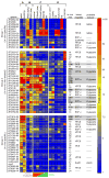
Figure 2. Responses of host glycan-induced PULs to fractionated PMG and purified glycans
A heat map showing PUL operon induction by various host glycans. Each box represents the average fold-change (relative to MM-glucose) of all genes within the indicated operon (gene numbers listed vertically at left), and is calibrated according to the vertical bar at the extreme right. Individual PULs are separated by horizontal breaks in the heat map. Only PUL operons with average fold-change values ≥10 are shown. The gene coordinates of several PULs listed in the text are more expansive. Growth conditions are separated into four groups: (columns A,B) the original in vivo and in vitro datasets, respectively, outlined in Fig. 1; (column C) PMG glycan fractions; and (column D) pure forms of various host glycans or disaccharide components. The mucin O-glycan and GAG distribution in fractionated PMG glycans is schematized at the bottom of column ‘C’ as a reference. Columns adjacent to the vertical scale bar on the right describe the following notable characteristics of each PUL/operon: ‘in vivo max’, the maximum induction observed in either in vivo dataset for Group 2 and Group 3 loci (gray bars are used to highlight PULs with operons showing maximum induction >100-fold); ‘linked regulator’, the type of regulator associated with each PUL; ‘probable inducer’, the class of host-glycan that appears to induce each PUL (dashes indicate the lack of a conspicuously associated regulator or an inability to clearly predict the type of inducing host glycan from the data). Abbreviations: N-acetyllactosamine (LacNAc), chondroitin sulfate (CS), heparin sulfate (HS), hybrid two-component system (HTCS); extracytoplasmic function sigma/anti-sigma pair (ECF-σ, homologs of the inner membrane-spanning maltose sensor SusR (SusR-like), and cytoplasmic one-component system (OCS).
Figure 3
Figure 3. GAG utilization PULs
The genomic architecture of two PULs required for GAG utilization. Enzymatic functions are labeled according to their assigned CAZy glycoside hydrolase (GH) and polysaccharide lyase (PL) family membership. The susC homolog, BT3332 was previously identified in a transposon screen for chondroitin utilization mutants and named csuF (Cheng et al., 1995), although the surrounding sequences were not reported. The inset shows a model depicting the putative cellular locations of GAG-utilizing functions, and is based on similarities to the prototypic Sus PUL (Fig. S2). Inferred locations of constitutive and HTCS-activated promoters are indicated above PUL genes. Omega (Ω) symbols below HTCS genes indicate sites of plasmid insertions to disrupt these genes.
Figure 4
Figure 4. Mucin O-glycan utilization PULs
Four loci induced during growth in the presence of mucin O-glycans, and a fifth in vivo-specific system (BT1617-22). Note the presence of an extra N-terminal trans-envelope signaling domain on one of the susC homologs associated with each system (Koebnik, 2005). The anti-σ factor associated with the BT4240-50 locus was assembled in the VPI-5482 genome as two divergent genes (BT4248-49) but undergoes a DNA inversion that positions BT4249 upstream of BT4248, resulting in an intact anti-σ factor gene (see Fig. S12 for details). Gray boxes indicate genes for which elevated basal transcription was observed (see Supplemental Data). Delta (Δ) symbols below ECF-σ genes indicate that they were targeted for in-frame deletions for in vivo competition experiments.
Figure 5
Figure 5. Specificity of ECF-σ-linked PULs for O-glycan components
(A) Representative core 2 mucin O-glycan structure. Two common disaccharides, core 1 and N-acetyllactosamine (LacNAc), are highlighted by orange- and green-shaded boxes, respectively. Monosaccharide symbols: GalNAc (open squares); Gal (closed circles); GlcNAc (closed squares); Fuc (open triangles); and NeuNAc (closed diamonds). (B) GeneChip-based induction values for genes in the BT4631-36 locus during growth on equimolar amounts of core 1 monosaccharides, core 1 disaccharide, LacNAc monosaccharides, and LacNAc disaccharide. Values are expressed as fold-difference relative to growth on MM-glucose. Color codes are the same in B–D. (C) Responses of genes in the BT2559-62 locus to growth on O-glycan mono- and disaccharide components. (D) Responses of genes in the BT4244-50 locus to growth on O-glycan mono- and disaccharide components. Note the differing substrate specificities for LacNAc and core 1 disaccharides. Values represent the mean±range of two biological replicates performed for each substrate.
Figure 6
Figure 6. Effects of multiple ECF-σ deletions on mucin O-glycan foraging, fitness and transmission in vivo.
(A) Expression of five ECF-σ-linked mucin O-glycan PULs in isogenic wild-type, mutant (Δ5ECF-σ) and complemented B. thetaiotaomicron strains. Fold-differences in expression are calibrated to the same scale as Fig. 2. Numbers to the right of the GeneChip-based heat map indicate the percent of wild-type expression observed in the Δ5ECF-σ and complement strains. Note that in Δ5ECF-σ cells, some PUL systems still exhibit substantial induction in vivo, indicating that other mechanisms are partially responsible for their activation. Likewise, not all systems are restored to wild-type levels by complementation. (B) Competitive colonization of adult germfree mice fed a standard plant glycan-rich diet by wild-type (black squares), Δ5ECF-σ (red triangles) and complemented strains (blue circles) (values plotted are mean ± one standard deviation). (C) Colonization of mice that were started on the same plant glycan-rich diet as in panel B, and then switched to a simple sugar diet on day 2, by wild-type (black squares), Δ5ECF-σ (red triangles) and complemented strains (blue circles). (D) Relative proportions of each competing strain in the input inoculum, mother, a co-housed adult female germfree control, and pups at various points in the natural transmission experiment. Values shown for the mother and adult female control are the average of samples taken at days −8, 0, 4, 11 and 15, or days 4, 11 and 15 relative to delivery, respectively. Values shown for the pups are the average of eight individually assayed animals at postnatal day 15. Error bars in panels B–D represent 1 standard deviation. Instances where a given strain’s population differs significantly from another strain(s) at the same time point are indicated with asterisks, (color-matched to the strain from which they differ). Later time points where the Δ5ECF-σ population differed significantly from its own 2d level (red asterisks) are also indicated in panel C (*, P≤0.05; **, P≤0.01 by Student’s t-test). For statistical comparisons in panel C, population values for the wild-type and mutant strains were normalized according to their initial abundance relative to Δ5ECF-σ at day 2.

Similar articles

Cited by

References

    1. An G, Wei B, Xia B, McDaniel JM, Ju T, Cummings RD, Braun J, Xia L. Increased susceptibility to colitis and colorectal tumors in mice lacking core 3-derived O-glycans. J Exp Med. 2007;204:1417–1429. - PMC - PubMed
    1. Bayer EA, Belaich JP, Shoham Y, Lamed R. The cellulosomes: multienzyme machines for degradation of plant cell wall polysaccharides. Ann Rev Microbiol. 2004;58:521–554. - PubMed
    1. Bjursell MK, Martens EC, Gordon JI. Functional genomic and metabolic studies of the adaptations of a prominent adult human gut symbiont, Bacteroides thetaiotaomicron, to the suckling period. J Biol Chem. 2006;281:36269–36279. - PubMed
    1. Braun V, Mahren S. Transmembrane transcriptional control (surface signalling) of the Escherichia coli Fec type. FEMS Microbiol Rev. 2005;29:673–684. - PubMed
    1. Cawley TN, Ballou CE. Identification of two Saccharomyces cerevisiae cell wall mannan chemotypes. J Bacteriol. 1972;111:690–695. - PMC - PubMed

Publication types