

Estrogen receptor-alpha immunoreactive neurons in the brainstem and spinal cord of the female rhesus monkey: species-specific characteristics
- PMID: 18996446
- PMCID: PMC4641676
- DOI: 10.1016/j.neuroscience.2008.10.017
Estrogen receptor-alpha immunoreactive neurons in the brainstem and spinal cord of the female rhesus monkey: species-specific characteristics
Abstract
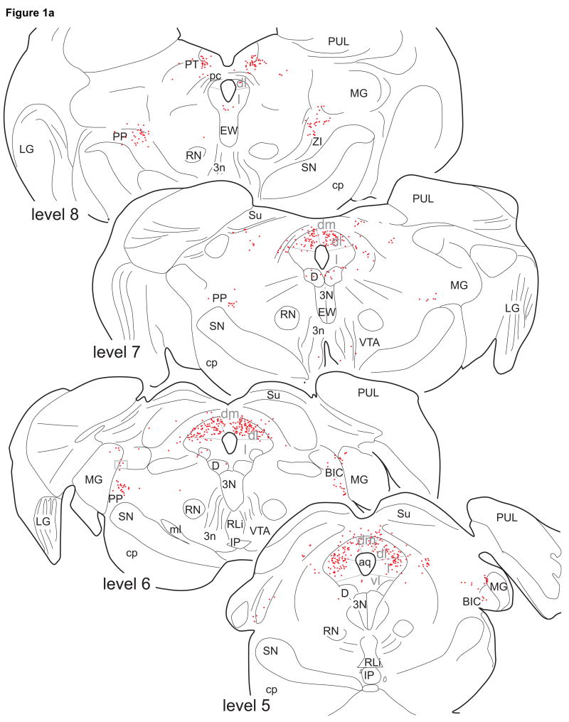
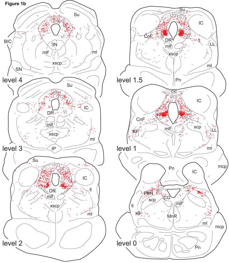
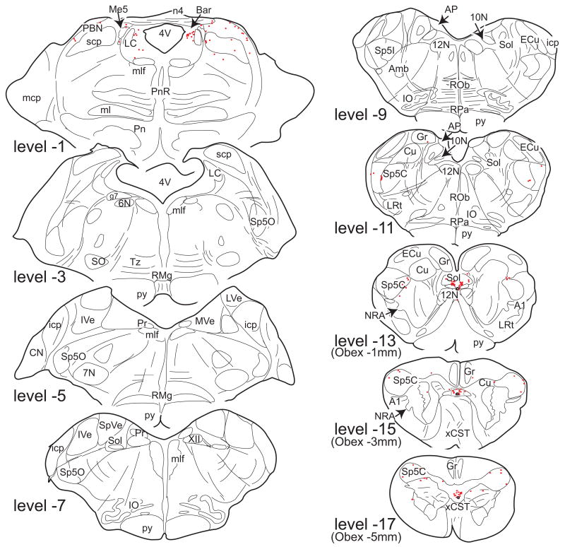
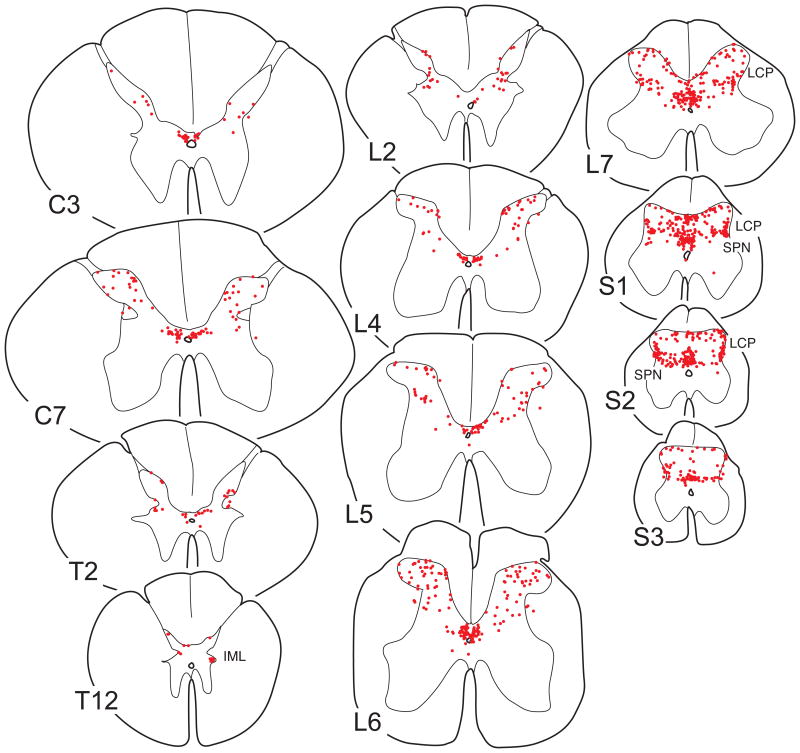
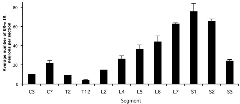
The distribution pattern of estrogen receptors in the rodent CNS has been reported extensively, but mapping of estrogen receptors in primates is incomplete. In this study we describe the distribution of estrogen receptor alpha immunoreactive (ER-alpha IR) neurons in the brainstem and spinal cord of the rhesus monkey. In the midbrain, ER-alpha IR neurons were located in the periaqueductal gray, especially the caudal ventrolateral part, the adjacent tegmentum, peripeduncular nucleus, and pretectal nucleus. A few ER-alpha IR neurons were found in the lateral parabrachial nucleus, lateral pontine tegmentum, and pontine gray medial to the locus coeruleus. At caudal medullary levels, ER-alpha IR neurons were present in the commissural nucleus of the solitary complex and the caudal spinal trigeminal nucleus. The remaining regions of the brainstem were devoid of ER-alpha IR neurons. Spinal ER-alpha IR neurons were found in laminae I-V, and area X, and were most numerous in lower lumbar and sacral segments. The lateral collateral pathway and dorsal commissural nuclei of the sacral cord and the thoracic intermediolateral cell column also contained ER-alpha IR neurons. Estrogen treatment did not result in any differences in the distribution pattern of ER-alpha IR neurons. The results indicate that ER-alpha IR neurons in the primate brainstem and spinal cord are concentrated mainly in regions involved in sensory and autonomic processing. Compared with rodent species, the regional distribution of ER-alpha IR neurons is less widespread, and ER-alpha IR neurons in regions such as the spinal dorsal horn and caudal spinal trigeminal nucleus appear to be less abundant. These distinctions suggest a modest role of ER-alpha in estrogen-mediated actions on primate brainstem and spinal systems. These differences may contribute to variations in behavioral effects of estrogen between primate and rodent species.
Figures

References
-
- Alves SE, Weiland NG, Hayashi S, McEwen BS. Immunocytochemical localization of nuclear estrogen receptors and progestin receptors within the rat dorsal raphe nucleus. J Comp Neurol. 1998;391:322–334. - PubMed
-
- Amandusson A, Hermanson O, Blomqvist A. Estrogen receptor-like immunoreactivity in the medullary and spinal dorsal horn of the female rat. Neurosci Lett. 1995;196:25–28. - PubMed
-
- An X, Bandler R, Ongur D, Price JL. Prefrontal cortical projections to longitudinal columns in the midbrain periaqueductal gray in macaque monkeys. J Comp Neurol. 1998;401:455–479. - PubMed
-
- Bandler R, Shipley MT. Columnar organization in the midbrain periaqueductal gray: modules for emotional expression? Trends Neurosci. 1994;17:379–389. - PubMed
Publication types
MeSH terms
Substances
Grants and funding
LinkOut - more resources
Full Text Sources
