

Functional brain correlates of social and nonsocial processes in autism spectrum disorders: an activation likelihood estimation meta-analysis
- PMID: 18996505
- PMCID: PMC2993772
- DOI: 10.1016/j.biopsych.2008.09.022
Functional brain correlates of social and nonsocial processes in autism spectrum disorders: an activation likelihood estimation meta-analysis
Abstract
Background: Functional neuroimaging studies of autism spectrum disorders (ASD) have examined social and nonsocial paradigms, although rarely in the same study. Here, we provide an objective, unbiased survey of functional brain abnormalities in ASD, related to both social and nonsocial processing.
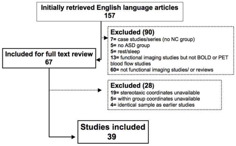
Methods: We conducted two separate voxel-wise activation likelihood estimation meta-analyses of 39 functional neuroimaging studies consisting of 24 studies examining social processes (e.g., theory of mind, face perception) and 15 studies examining nonsocial processes (e.g., attention control, working memory). Voxel-wise significance threshold was p<.05, corrected by false discovery rate.
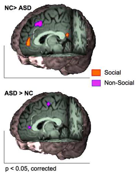
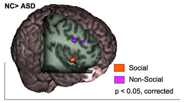
Results: Compared with neurotypical control (NC) subjects, ASD showed greater likelihood of hypoactivation in two medial wall regions: perigenual anterior cingulate cortex (ACC) in social tasks only and dorsal ACC in nonsocial studies. Further, right anterior insula, recently linked to social cognition, was more likely to be hypoactivated in ASD in the analyses of social studies. In nonsocial studies, group comparisons showed greater likelihood of activation for the ASD group in the rostral ACC region that is typically suppressed during attentionally demanding tasks.
Conclusions: Despite substantial heterogeneity of tasks, the rapidly increasing functional imaging literature showed ASD-related patterns of hypofunction and aberrant activation that depended on the specific cognitive domain, i.e., social versus nonsocial. These results provide a basis for targeted extensions of these findings with younger subjects and a range of paradigms, including analyses of default mode network regulation in ASD.
Conflict of interest statement
Figures
References
-
- Volkmar FR, Lord C, Bailey A, Schultz RT, Klin A. Autism and pervasive developmental disorders. J Child Psychol Psychiatry. 2004;45:135–170. - PubMed
-
- Schultz RT. Developmental deficits in social perception in autism: the role of the amygdala and fusiform face area. Int J Dev Neurosci. 2005;23:125–141. - PubMed
-
- Frith U. Mind blindness and the brain in autism. Neuron. 2001;32:969–979. - PubMed
-
- Klin A, Sparrow SS, de Bildt A, Cicchetti DV, Cohen DJ, Volkmar FR. A normed study of face recognition in autism and related disorders. J Autism Dev Disord. 1999;29:499–508. - PubMed
-
- Klin A, Jones W, Schultz R, Volkmar F, Cohen D. Visual fixation patterns during viewing of naturalistic social situations as predictors of social competence in individuals with autism. Archives of General Psychiatry. 2002;59:809–816. - PubMed
Publication types
MeSH terms
Grants and funding
LinkOut - more resources
Full Text Sources
