Transperineal three-dimensional ultrasound imaging for detection of anatomic defects in the anal sphincter complex muscles
- PMID: 18996750
- PMCID: PMC2835972
- DOI: 10.1016/j.cgh.2008.08.019
Transperineal three-dimensional ultrasound imaging for detection of anatomic defects in the anal sphincter complex muscles
Abstract
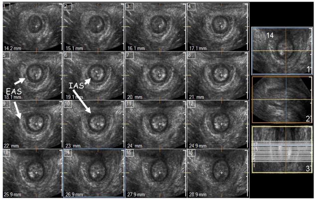
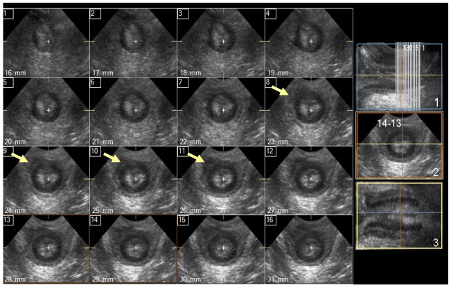
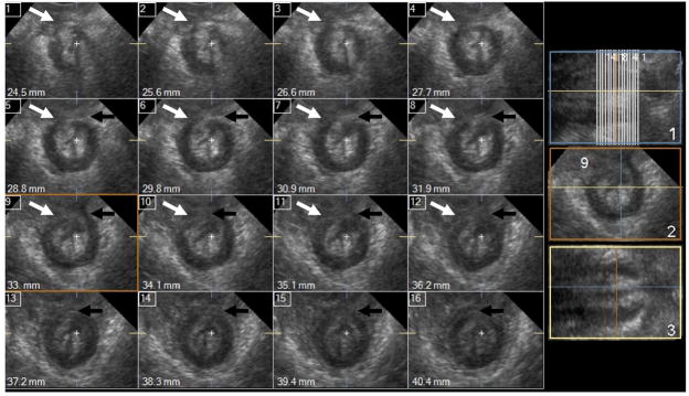
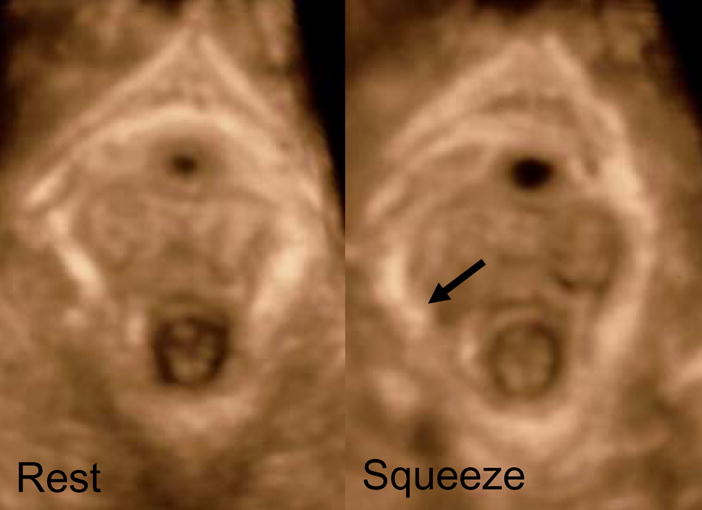
Background & aims: Three-dimensional ultrasound (3D-US) imaging is a powerful tool to visualize various components of the anal sphincter complex, that is, the internal anal sphincter (IAS), the external anal sphincter (EAS), and the puborectalis muscle (PRM). Our goal was to determine the reliability of the 3D-US imaging technique in detecting morphologic defects in the IAS, EAS, and PRM.
Methods: Transperineal 3D-US images were obtained in 3 groups of women: nulliparous (n = 13), asymptomatic parous (n = 20), and patients with fecal incontinence (FI) (n = 25). The IAS and EAS were assessed to determine the craniocaudal length of defects and were scored as follows: 0 = normal, 1 = less than 25%, 2 = 25% to 50%, 3 = 50% to 75%, and 4 = greater than 75%. The 2 PRM hemislings were scored separately as follows: 0 = normal, 1 = less than 50% abnormal, and 2 = greater than 50% length abnormal. Subjects were grouped according to the score as follows: normal (score 0), minor abnormality (scores of 1 and 2), and major abnormality (scores of 3 and 4). Three observers performed the scoring.
Results: The 3D-US allowed detailed evaluation of the IAS, EAS, and PRM. The inter-rater reliability for detecting the defects ranged between 0.80 and 0.95. Nullipara women did not show any significant defect but the defects were quite common in asymptomatic parous and FI patients. The prevalence of defects was greater in the FI patients as compared with the asymptomatic parous women.
Conclusions: 3D-US yields reliable assessment of morphologic defects in the anal sphincter complex muscles.
Conflict of interest statement
Figures


Similar articles
-
Anal sphincter defects and fecal incontinence 15-24 years after first delivery: a cross-sectional study.Ultrasound Obstet Gynecol. 2018 May;51(5):677-683. doi: 10.1002/uog.18827. Ultrasound Obstet Gynecol. 2018. PMID: 28782264
-
Anal sphincter complex muscles defects and dysfunction in asymptomatic parous women.Int Urogynecol J. 2011 Sep;22(9):1143-50. doi: 10.1007/s00192-011-1446-8. Epub 2011 May 21. Int Urogynecol J. 2011. PMID: 21604060
-
Functional morphology of anal sphincter complex unveiled by high definition anal manometery and three dimensional ultrasound imaging.Neurogastroenterol Motil. 2011 Nov;23(11):1013-9, e460. doi: 10.1111/j.1365-2982.2011.01782.x. Epub 2011 Sep 25. Neurogastroenterol Motil. 2011. PMID: 21951657 Free PMC article.
-
Transperineal three-dimensional ultrasound in evaluating anal sphincter muscles.Ultrasound Obstet Gynecol. 2007 Aug;30(2):201-9. doi: 10.1002/uog.4057. Ultrasound Obstet Gynecol. 2007. PMID: 17605148
-
Diagnostic capabilities of transperineal ultrasound (TPUS) to evaluate anal sphincter defect post obstetric anal sphincter injury (OASIS)? A systematic review.J Ultrasound. 2023 Jun;26(2):393-399. doi: 10.1007/s40477-022-00763-3. Epub 2023 Jan 11. J Ultrasound. 2023. PMID: 36630015 Free PMC article.
Cited by
-
Current applications of transperineal ultrasound in gastroenterology.World J Radiol. 2016 Apr 28;8(4):370-7. doi: 10.4329/wjr.v8.i4.370. World J Radiol. 2016. PMID: 27158423 Free PMC article. Review.
-
The evolution of transperineal ultrasound findings of the external anal sphincter during the first years after childbirth.Int Urogynecol J. 2016 Dec;27(12):1899-1903. doi: 10.1007/s00192-016-3055-z. Epub 2016 Jun 1. Int Urogynecol J. 2016. PMID: 27250832
-
Purse-string morphology of external anal sphincter revealed by novel imaging techniques.Am J Physiol Gastrointest Liver Physiol. 2014 Mar;306(6):G505-14. doi: 10.1152/ajpgi.00338.2013. Epub 2014 Jan 23. Am J Physiol Gastrointest Liver Physiol. 2014. PMID: 24458022 Free PMC article.
-
Anatomical disruption and length-tension dysfunction of anal sphincter complex muscles in women with fecal incontinence.Dis Colon Rectum. 2013 Nov;56(11):1282-9. doi: 10.1097/DCR.0b013e3182a18e87. Dis Colon Rectum. 2013. PMID: 24105004 Free PMC article.
-
Anal Sphincter Anatomy Prepregnancy to Postdelivery Among the Same Primiparous Women on Dynamic Magnetic Resonance Imaging.Female Pelvic Med Reconstr Surg. 2019 Jan/Feb;25(1):8-14. doi: 10.1097/SPV.0000000000000504. Female Pelvic Med Reconstr Surg. 2019. PMID: 29068801 Free PMC article.
References
-
- Sultan AH, Kamm MA, Hudson CN, Thomas JM, Bartram CI. Anal-sphincter disruption during vaginal delivery. N Engl J Med. 1993;329(26):1905–11. - PubMed
-
- Fernandez-Fraga X, Azpiroz F, Malagelada JR. Significance of pelvic floor muscles in anal incontinence. Gastroenterology. 2002;123(5):1441–50. - PubMed
-
- Bharucha AE. Fecal incontinence. Gastroenterology. 2003;124(6):1672–85. - PubMed
-
- DeLancey JO. The anatomy of the pelvic floor. Curr Opin Obstet Gynecol. 1994;6(4):313–6. - PubMed
Publication types
MeSH terms
Grants and funding
LinkOut - more resources
Full Text Sources