. 2009 Jan 9;284(2):966-73.
doi: 10.1074/jbc.M806830200. Epub 2008 Nov 6.

ABF1-binding sites promote efficient global genome nucleotide excision repair

Affiliations

ABF1-binding sites promote efficient global genome nucleotide excision repair

Shirong Yu et al. J Biol Chem. .

Abstract

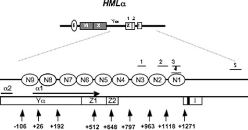
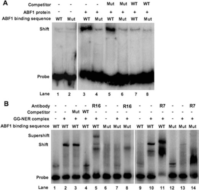
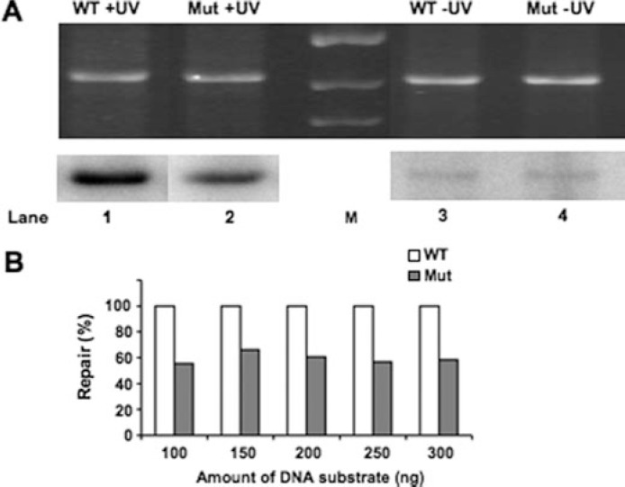
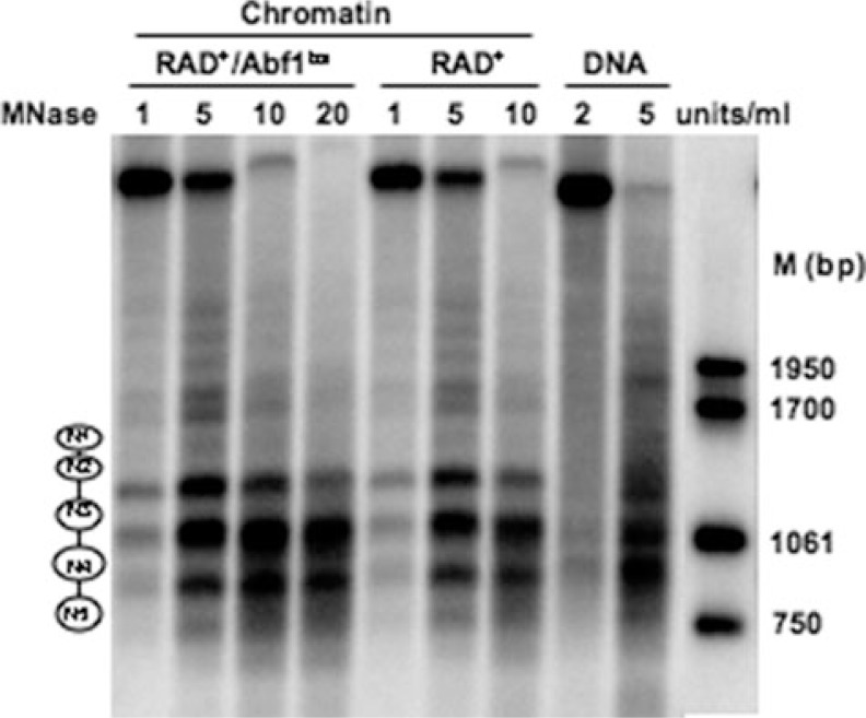
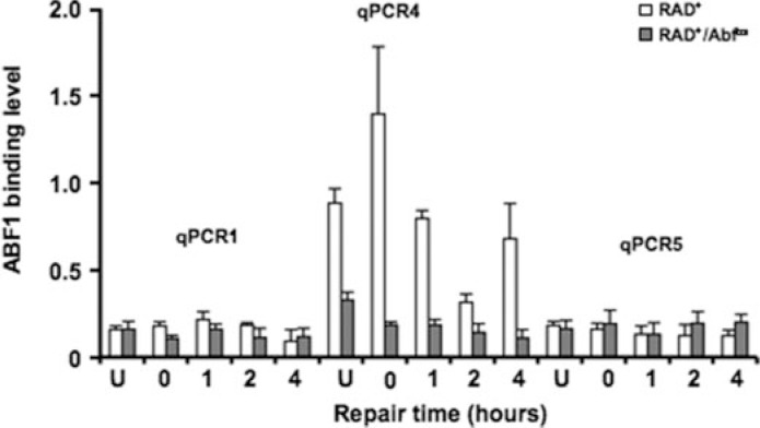
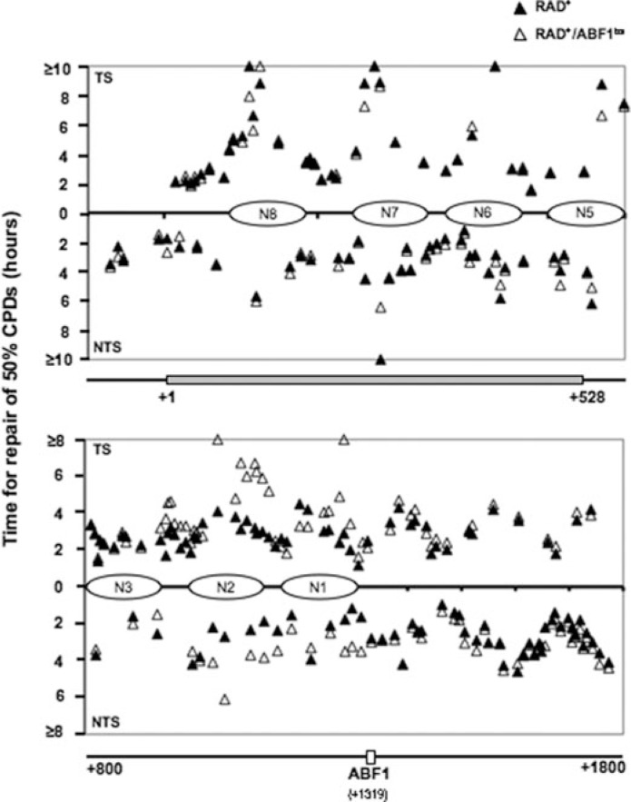
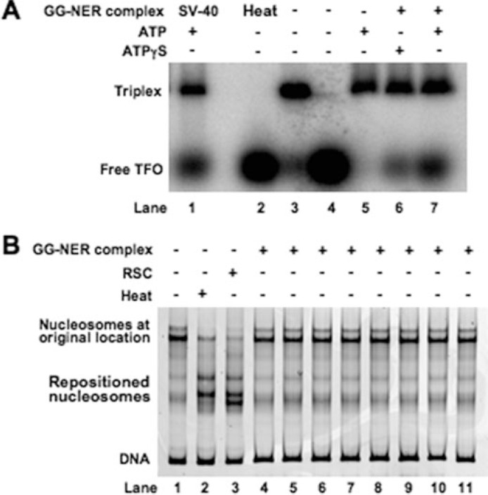
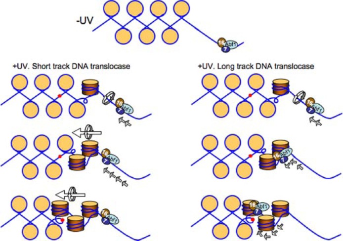
Global genome nucleotide excision repair (GG-NER) removes DNA damage from nontranscribing DNA. In Saccharomyces cerevisiae, the RAD7 and RAD16 genes are specifically required for GG-NER. We have reported that autonomously replicating sequence-binding factor 1 (ABF1) protein forms a stable complex with Rad7 and Rad16 proteins. ABF1 functions in transcription, replication, gene silencing, and NER in yeast. Here we show that binding of ABF1 to its DNA recognition sequence found at multiple genomic locations promotes efficient GG-NER in yeast. Mutation of the I silencer ABF1-binding site at the HMLalpha locus caused loss of ABF1 binding, which resulted in a domain of reduced GG-NER efficiency on one side of the ABF1-binding site. During GG-NER, nucleosome positioning at this site was not altered, and this correlated with an inability of the GG-NER complex to reposition nucleosomes in vitro.We discuss how the GG-NER complex might facilitate GG-NER while preventing unregulated gene transcription during this process.


Figures

FIGURE 1.
FIGURE 1.
Schematic representation of the physical map of HMLα locus (adapted from Weiss and Simpson (25)). The I silencer is indicated by a white box labeled “I.” The ABF1 binding sequence is shown by a black bar; open ellipses indicate positioned nucleosomes (N1–N9). The α1 coding region is identified by the horizontal arrow. The numbered vertical arrows mark the relative nucleotide positions from the α1 ATG start codon; A is +1. qPCR regions (1–5) are as follows: qPCR1, +871 to +959; qPCR2, +1056 to +1156; qPCR3, +1163 to +1248; qPCR4, +1162 to +1278; qPCR5, +1884 to +2095.
FIGURE 2.
FIGURE 2.
EMSAs to detect ABF1 and GG-NER complex DNA binding activity. A, EMSAs with ABF1. [γ-32P]dATP-labeled 480-bp duplex DNA containing wild-type ABF1 binding sequence (WT; lanes 1, 3, 5, and 7) or triple point-mutated ABF1 binding sequence (Mut; lanes 2, 4, 6, and 8) was incubated at room temperature for 30 min either with (lanes 3– 8) or without recombinant ABF1 (lanes 1 and 2). A competition experiment was performed in the presence of unlabeled DNAs containing three point-mutated (lanes 5 and 6) or wild-type (lanes 7 and 8) ABF1 binding sequence. B, EMSAs with GG-NER complex. [γ-32P]dATP-labeled 480-bp duplex DNA containing wild-type ABF1 binding sequence (lanes 1–5 and 9–11) or triple point-mutated ABF1 binding sequence (Mut; lanes 6–8 and 12–14) was incubated at room temperature for 30 min either with (lanes 2–5, 7, 8, 10, 11, 13, and 14) or without GG-NER complex (lanes 1, 6, 9, and 12). Competition was performed in the presence of unlabeled DNAs containing triple point-mutated (Mut; lane 3) and normal (WT; lane 4) ABF1 binding sequence. In supershift assays, antibodies against Rad16 (R16) (lanes 5 and 8) and Rad7 (R7) (lane 11 and 14) were subsequently added to the mixture and incubated for another 30 min at room temperature.
FIGURE 3.
FIGURE 3.
In vitro NER analysis to monitor repair synthesis. NER was performed at 26 °C for 2 h using UV radiation-damaged (+UV; 300 J/m2) or undamaged (−UV) plasmid pUC18-ABF1-HIS3 (WT; lanes 1 and 3, respectively) or pUC18-ABF1bs-HIS3 (Mut; lanes 2 and 4, respectively) in the presence of wild-type yeast whole cell extract. A, ethidium bromide-stained gel represents the total DNA loading (top), and an autoradiograph of the gel indicates the level of DNA repair synthesis or background radiolabel incorporation for the damaged (+UV) or undamaged (−UV) plasmids (bottom). M, DNA marker lane. B, relative levels of DNA repair radiolabel incorporation for the damaged (+UV) or undamaged (−UV) plasmids at the range of DNA substrate amounts indicated.
FIGURE 4.
FIGURE 4.
Low resolution micrococcal nuclease nucleosome mapping. The amount of micrococcal nuclease (MNase) used is indicated at the top of each lane. Molecular weight markers (M) on the right indicate the size (bp) of the bands. The positioned nucleosomes (N1–N5) are shown as ellipses on the left.
FIGURE 5.
FIGURE 5.
ABF1 binding to HMLα I silencer requires an ABF1-binding site. Immunoprecipitation with ABF1 antibody was performed in RAD+ and RAD+/Abf1bs cells. The occupancy of ABF1 was measured by qPCR with the primers spanning the HMLα I silencer region (qPCR1, qPCR4, and qPCR5). Data are represented as average of three experiments ±S.D.
FIGURE 6.
FIGURE 6.
CPD repair in RAD+ and RAD+/ABF1bs strains. The gels (supplemental Figs. 4–6) were quantified with ImageQuant software. Repair time for 50% of CPDs (t50%) at a site was calculated or extrapolated. Closed triangles represent CPD repair in the RAD+ strain; open triangles represent CPD repair in the RAD+/ABF1bs. Horizontal bars at the bottom of each graph show regions examined, and numbers indicate nucleotide positions.
FIGURE 7.
FIGURE 7.
DNA translocase and nucleosome sliding activity of GG-NER complex. A, triple helix substrates consisting of a 40-nucleotide triple helical region were prepared as described under “Experimental Procedures.” Substrates were incubated with (lanes 6–7) or without (lane 3) GG-NER complex at 30 °C for 30 min in the presence of ATP (lanes 5 and 7) or ATPγS (lane 6) or heated for 5 min at 90 °C (lane 2). SV-40 T-antigen-treated sample (lane 1) is included as a positive control. Lane 4 contains [γ-32P]dATP-labeled third strand (indicated as Free TFO) only. A control fraction from the final step of the purification known not to contain GG-NER complex was also tested (lane 5). Reaction mixtures were separated on a 1% agarose gel, and displacement of the free triplex-forming oligonucleotide (TFO) was monitored. B, movement of nucleosomes on the Cy3-labeled DNA fragment 105A64 was monitored following heating at 47 °C for 1 h (lane 2) or treatment with different GG-NER complex-containing fractions from the final step of the purification as indicated previously (16) (lanes 4–11) or 0.12 pmol of RSC complex (lane 3) for 30 min by native gel electrophoresis.
FIGURE 8.
FIGURE 8.
Proposed model of GG-NER complex function at the HMLα locus ABF1-binding site. In the absence of UV radiation the GG-NER complex binds in a specific orientation to the ABF1-binding site (top panel). Following UV radiation the directional DNA translocase activity of the complex results in changes in occupancy of the GG-NER complex at the ABF1-binding site. This generates a domain of altered superhelical torsion in one direction from the ABF1-binding site. This action promotes efficient GG-NER within the domain. The GG-NER complex could generate superhelical torsion by DNA translocation over a short distance from the ABF1-binding site, for example tens of base pairs generating a domain of torsion over hundreds of base pairs (left panel), or by long distance tracking of the complex throughout the domain (right panel). Note that the translational setting of the nucleosomes (beige circles) on the DNA is not perturbed by this process. DNA damage is represented by red spots. 7, Rad7; 16, Rad16.

Similar articles

Cited by

References

    1. Diffley JF, Stillman B. Science. 1989;246:1034–1038. - PubMed
    1. Reid JL, Iyer VR, Brown PO, Struhl K. Mol. Cell. 2000;6:1297–1307. - PubMed
    1. Yarragudi A, Miyake T, Li R, Morse RH. Mol Cell Biol. 2004;24:9152–9164. - PMC - PubMed
    1. Rao H, Stillman B. Proc Natl Acad Sci U S A. 1995;92:2224–2228. - PMC - PubMed
    1. Zou Y, Yu Q, Bi X. Mol Cell Biol. 2006;26:7806–7819. - PMC - PubMed

Publication types

MeSH terms