. 2009 Feb 1;18(3):497-516.
doi: 10.1093/hmg/ddn377. Epub 2008 Nov 7.

Disruption of neural progenitors along the ventricular and subventricular zones in periventricular heterotopia

Affiliations

Disruption of neural progenitors along the ventricular and subventricular zones in periventricular heterotopia

Russell J Ferland et al. Hum Mol Genet. .

Abstract

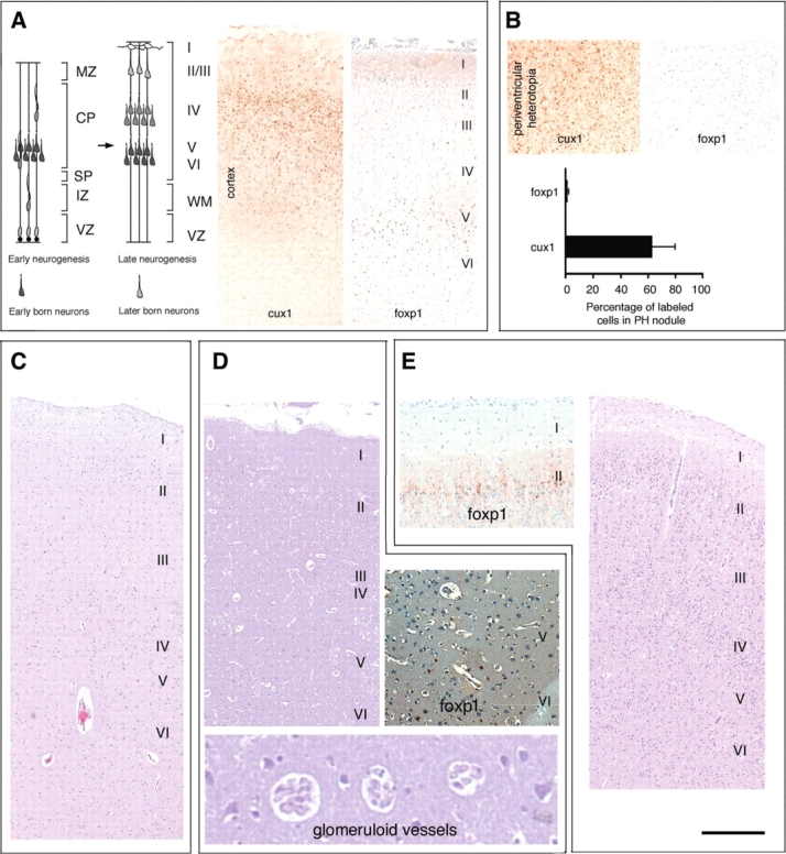
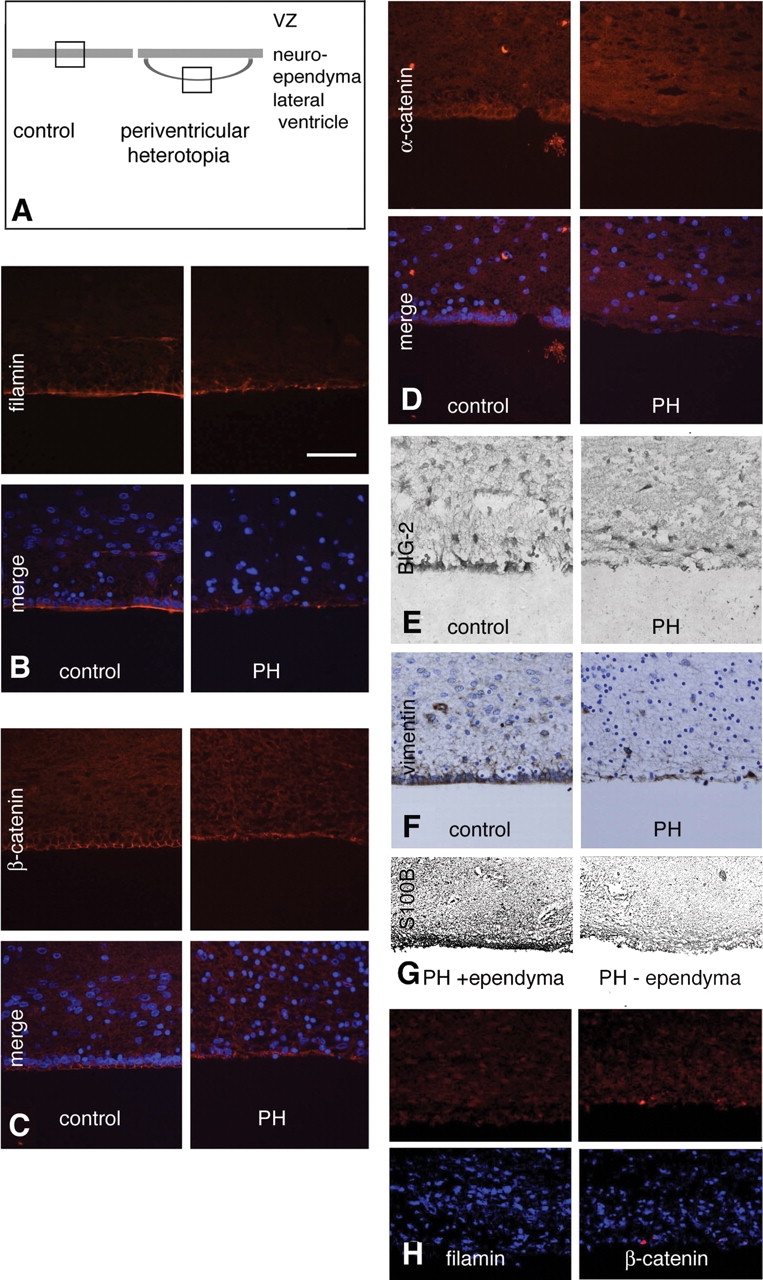
Periventricular heterotopia (PH) is a disorder characterized by neuronal nodules, ectopically positioned along the lateral ventricles of the cerebral cortex. Mutations in either of two human genes, Filamin A (FLNA) or ADP-ribosylation factor guanine exchange factor 2 (ARFGEF2), cause PH (Fox et al. in 'Mutations in filamin 1 prevent migration of cerebral cortical neurons in human periventricular heterotopia'. Neuron, 21, 1315-1325, 1998; Sheen et al. in 'Mutations in ARFGEF2 implicate vesicle trafficking in neural progenitor proliferation and migration in the human cerebral cortex'. Nat. Genet., 36, 69-76, 2004). Recent studies have shown that mutations in mitogen-activated protein kinase kinase kinase-4 (Mekk4), an indirect interactor with FlnA, also lead to periventricular nodule formation in mice (Sarkisian et al. in 'MEKK4 signaling regulates filamin expression and neuronal migration'. Neuron, 52, 789-801, 2006). Here we show that neurons in post-mortem human PH brains migrated appropriately into the cortex, that periventricular nodules were primarily composed of later-born neurons, and that the neuroependyma was disrupted in all PH cases. As studied in the mouse, loss of FlnA or Big2 function in neural precursors impaired neuronal migration from the germinal zone, disrupted cell adhesion and compromised neuroepithelial integrity. Finally, the hydrocephalus with hop gait (hyh) mouse, which harbors a mutation in Napa [encoding N-ethylmaleimide-sensitive factor attachment protein alpha (alpha-SNAP)], also develops a progressive denudation of the neuroepithelium, leading to periventricular nodule formation. Previous studies have shown that Arfgef2 and Napa direct vesicle trafficking and fusion, whereas FlnA associates dynamically with the Golgi membranes during budding and trafficking of transport vesicles. Our current findings suggest that PH formation arises from a final common pathway involving disruption of vesicle trafficking, leading to impaired cell adhesion and loss of neuroependymal integrity.


Figures

Figure 1.
Figure 1.
Histopathology in human periventricular heterotopia (PH). (A) T2-weighted MR image of the brain from a 2-month-old female with PH (Case 3). Small gray matter nodules of neurons (white arrowheads) can be seen lining the lateral ventricles. (B) Gross specimen of the same case demonstrates a periventricular nodule (black arrow) with otherwise normal appearing cortex and basal ganglia. (C) Hemotoxylin–eosin staining shows a normal six-layered cortex and contiguous nodules (asterisks) along the lateral ventricle (higher magnification to the right). (D) Occasional regions of cortical dysplasia are seen with larger pyramidal neurons ectopically located in the more superficial layers of the cortex. However, the vast majority of the cortical layers appear normal. (E) The nodule is comprised of neurons, expressing a variety of neuronal-specific markers.
Figure 2.
Figure 2.
Neurons expressing the mutant FLNA protein migrate appropriately into the cerebral cortex. (A) Photomicrographs of the cerebral cortex and the nodular heterotopia from the 2-month-old female, immunostained with the superficial layer marker (CUX1) and the deep-layer marker (FOXP1), demonstrate a normal cortical lamination with no blurring of the cortical layer boundaries. (B) Photomicrograph of the heterotopic nodules in the 2-month-old female demonstrate increased numbers of CUX1-positive neurons, as compared to FOXP1-positive neurons, suggesting that these nodules are composed of later-born neurons. (C) Bright field photomicrograph of the cortex of an 83-year-old male with PH. The cortical layers appear discrete with no blurring of laminae on Nissl stain. (D) Nissl stained cortical section of a 27-year-old female similarly shows preservation of the layers. Immunostaining for FOXP1, which labels the deep-layer neurons, shows positive and appropriate staining in laminae V/VI. Glomeroid vascular anomalies were also seen in the cortex and have previously been reported in PH cases due to FLNA mutations. (E) Nissl stained cortical section of a 37-week GA male derived from a familial PH pedigree with a previously reported FLNA mutation shows appropriate cortical layering by Nissl stain. Positive immunostaining for FOXP1 which in embryonic human brain labels the neurons of the superficial layers (45).
Figure 3.
Figure 3.
Disruption of the neuroependyma lining along the periventricular nodules in PH brains. (A) Schematic demonstrates the region of impaired neuroepithelial integrity in the neuronal nodules. (BF) Photomicrographs of tissues from control and PH cases (2-month-old female with PH), following immunostaining for FLNA (B), β-catenin (C), α-catenin (D), BIG-2 (E), vimentin (F) and S100B (G) shows loss of the neuroependymal lining along the heterotopia. Moreover, in areas within PH brains, where the ependyma is intact, there is no disruption of radial glial differentiation as assessed by S100B staining (G). Finally, both FLNA and BIG2 (the protein encoding the gene shown to cause the autosomal recessive form of PH) (37) expression patterns are notably confined to the neuroepithelial lining (B and E). (H) Fluorescent photomicrographs of FLNA and β-catenin staining along the neuroependymal lining of the ventricular zone in a 37-week-old GA male with PH and a FLNA mutation demonstrating the disruption along the ventricular zone.
Figure 4.
Figure 4.
Loss of FlnA function disrupts neuroepithelial cell contacts along the murine ventricular lining. (A) The neuroependymal lining (arrowheads), as assessed by β-catenin staining, appears disrupted 48 h after in utero electroporation of the dominant-negative EGFP-ΔABD-FilaminA construct, by comparison to an EGFP control construct, via confocal microscopy. Electroporation is performed on embryonic day 16.5 (E16.5) embryos and immunostaining is performed in the E18.5 embryo. The number of GFP-positive cells along the lining with co-localized surface expression of β-catenin is quantified relative to the total number of GFP-positive cells, following transfection of either control or EGFP-ΔABD-FilaminA construct. This change in β-catenin distribution and alteration in cell contacts are quantified below (Student's t-test: **P < 0.02; control n = 10, experimental n = 5). (B) The neuroependymal lining, as assessed by α-catenin, is also disrupted 48 h after in utero electroporation of the dominant-negative EGFP-ΔABD-FilaminA construct. Electroporation is performed on E16.5 embryos and immunostaining is performed in the E18.5 embryo. (C) MDCK cell cultures transfected with the dominant-negative EGFP-ΔABD-FilaminA similarly leads to a partial loss of cell–cell contacts (β-catenin, small arrowheads) and alterations in β-catenin distribution as compared to cells transfected with EGFP alone. Cultures are maintained for 48 h after transfection prior to immunostaining with the cell adhesion marker, β-catenin. The number of transfected cells with disrupted cell surface expression of β-catenin was quantified in both control and EGFP-ΔABD-FilaminA positive cells. The alterations in cell contacts are summarized to the right (Student's t-test: **P < 0.02; control n ≥ 3, experimental n ≥ 3 independent experiments). (D) Fluorescent photomicrograph of neural precursors 2 days after transfection either with control EGFP or dominant-negative EGFP-ΔABD-FilaminA constructs, followed by staining for the actin cytoskeleton with phalloidin (rhodamine). Inhibition of FLNA leads to a more rounded appearance in the precursor cells and overall loss in cell membrane spreading, consistent with a disruption in cell adhesion. Cell areas of transfected cells are measured using NIH ImageJ software (Student's t-test: **P < 0.05; control n ≥ 3, experimental n ≥ 3 independent experiments). (E) Fluorescent photomicrograph of neural precursors 2 days after transfection with control EGFP or dominant-negative EGFP-ΔABD-FilaminA constructs, followed by staining for the apoptotic cell death marker caspase3. The rounded appearance of the neural cells following inhibition of FLNA is not due to increased cell death (scale bar = 25 µm).
Figure 5.
Figure 5.
Inhibition of Arfgef2/Big2 interrupts the integrity of the neuroependymal lining and leads to periventricular nodule formation in mice. (A) Periventricular heterotopic nodules (PH1 and PH2) and enlarged ventricles (LV) are seen on Cresyl violet stained tissue in a 2-week-old mouse following early post-natal intraventricular injections of 40 µm BFA. Heterotopic nodules (PH1) were seen just below the ventricular surface. Clusters of cells (PH2, arrowheads) also extended beyond the neuroependymal lining to lie directly along the LV. Immunostaining for these ectopically localized cells shows that they express the neuronal marker NeuN (below). To the right, the higher magnification photomicrographs of PH1 and PH2 show neurons ectopically positioned along the ventricle. (B) Two hours after intraventricular injection of BFA into E16.5 mice, phalloidin and N-cadherin staining along the ventricular zone neuroependyma is discontinuous. (C) Within 7 h after intraventricular injection into E16.5 mice, BFA disrupts the continuity of β-catenin staining along the ventricular neuroepithelium. (D) Through inhibition of Big2, BFA impairs transport of adhesion molecules such as β-catenin from the Golgi apparatus to the cell surface in MDCK cells. This impairment in vesicle transport likely contributes to heterotopia formation. (E) After exposure to BFA for 7 h, the trafficking of β-catenin from the Golgi complex to the cell membrane is disrupted.
Figure 6.
Figure 6.
Loss of alpha-SNAP function in hyh mice disrupts the integrity of the neuroepithelium (ventricular zone). Coronal sections, stained with hematoxylin-eosin, through the brain of wild-type (wt) and mutant hyh (hyh) mice, at E12.5, E14.5 and E16.5. A caudal (fourth ventricle) to rostral (lateral ventricles) progressive disruption of the ventricular zone (VZ) lining the ventricular system is shown. (A and B) At E12.5, the neuroepithelium of the ganglionic eminence (GE) appears intact, while neuroepithelium denudation has already started at the fourth ventricle in the hyh mouse (inserts in lower left and right corners). (C and D) At E14.5, loss of neuroepithelium occurs along the caudal GE in the hyh mouse (arrows, compare inset images in C and D). (E and F) By E16.5, a breakdown of neuroepithelium and disorganization of the subventricular zone (SVZ) reaches the rostral horns of lateral ventricles in the hyh mouse (arrows, compare inset images in E and F). In the hyh mouse, denudation of the neuroepithelium leads to protrusion of progenitor cells from the SVZ into the ventricular lumen (top inset imagess in D and F). (GJ) Immunolocalization of alpha-SNAP in the GE in wild-type tissue at E14.5 (G–H) and the cerebral cortex at E16.5 (I–J). (G and I) Alpha-SNAP immunoreactivity is mainly found in the cells of the VZ (VZ, arrows). (H and J) Differential interference contrast (DIC, Nomarski optics) imaging of the same area is shown in G and I, respectively. LV, lateral ventricle. Scale bars: 250 µm (A–F); 500 µm (low magnification insets in A–F); 50 µm (high magnification insets in A–F and G–J).
Figure 7.
Figure 7.
Loss of alpha-SNAP function in hyh mice disrupts the progenitor cell population along the neuroependyma. (AD) Wild-type mice; (EH) hyh mutant mice. (A) Sagittal, hematoxylin–eosin stained section through the CNS of an E14.5 wild-type mouse. Boxed area is shown in B. (B) Higher magnification image of the area of the ganglionic eminence (GE) shown in A. The integrity of the ventricular zone (VZ)/neuroepithelium and subventricular zone (SVZ) is clearly seen. Inset: caveolin-1, functional marker, also reveals the integrity of the VZ (arrow). (C) Section adjacent to that shown in (B), immunostained for β-III-tubulin. The cells of the VZ are not immunoreactive, while those of the SVZ express the neuronal marker. (D) Scanning electron micrograph of the ventricular surface of the GE of a wild-type E18.5 embryo, showing a mosaic-like arrangement of cells, most of which are monociliated (stem cells/radial glia) (arrows) with few multiciliated cells (immature ependyma). (E) Sagittal hematoxylin–eosin stained section through the CNS of a mutant hyh E14.5 mouse. Boxed area framed is shown in (F). (F) Higher-magnification image of the area of the GE shown in (F). Denudation of the neuroepithelium and exposure of the cells of the SVZ to the ventricular lumen are evident (arrows). A few patches of VZ still remain intact. Inset: Disorganization of the VZ and SVZ (asterisk) can also be shown by immunostaining for caveolin-1. Arrow points to a patch of VZ. (G) Section adjacent to that shown in (F), immunostained for β-III-tubulin. Immunoreactive cells of the SVZ are fully exposed to the ventricular cavity (arrows). VZ indicates an undetached patch of neuroepithelium. (H) Scanning electron micrograph of the ventricular surface of the GE of a mutant hyh (E18.5) embryo, revealing denudation of the neuroepithelium with progenitor cells (PC) lining the VZ with associated macrophages (M). Scale bars: 500 µm (A and E); 50 µm (B, D, F and G); 25 µm (insets in B and F); 10 µm (D and H).
Figure 8.
Figure 8.
Loss of alpha-SNAP function in hyh mice leads to PH formation. (A) Hematoxylin-eosin (H/E) stained coronal section through the brain of PN14 (P14) hyh mutant mouse, displaying nodular periventricular heterotopias (arrowheads) below the ependymal-denuded ventricular walls. Boxed area is shown in (C). CC, cerebral cortex; HI, hippocampus; LV, lateral ventricle. (B) Adjacent section to that of (A) immunostained for GLUT-1. The ependyma lining the hippocampus (HI) is preserved and GLUT-1 immunoreactive (arrow). The dorsal and lateral walls of the ventricle are denuded (asterisks). (C) Higher-magnification image of the area boxed in (A). Nodular periventricular heterotopias (PH1 and PH2, oval regions delineated by dashed lines) are seen adjacent to the ependymal-denuded ventricular wall (D and E) and the subventricular zone of the lateral wall. (E) Periventricular nodules are formed by cells with large nuclei and prominent nucleoli. The same section shown in (A, C was bleached and used for toluidine staining (E), bleached again and used for immunostaining (D). The two rectangular areas in C are shown in (D) and (E), respectively. (D) Immunostaining with a neuronal marker (β-III-tubulin) shows that the cells forming the periventricular nodules (PH1) in C are indeed neurons (arrows). (E) Similarly, toluidine blue staining demonstrates that periventricular nodules (PH2) in (C) displays a neuronal phenotype. (F) Frontal section through the brain of a hyh mutant mouse injected with BrdU from P4 (PN4) to P6 (PN6) and sacrificed 3 h after the last BrdU injection on P6. PH, periventricular heterotopia; SVZ, subventricular zone; CC, cerebral cortex. (G) Higher-magnification image of a portion of (F) showing that cells within the nodule (PH) do not proliferate post-natally, while those of the SVZ continue to proliferate post-natally and are BrdU-labeled (inset). Scale bars: 200 µm (A, B and F); 50 µm (C and G); 10 µm (D, E, inset in G).

Similar articles

Cited by

References

    1. Barkovich A.J., Koch T.K., Carrol C.L. The spectrum of lissencephaly: report of ten patients analyzed by magnetic resonance imaging. Ann. Neurol. 1991;30:139–146. - PubMed
    1. Dobyns W.B., Reiner O., Carrozzo R., Ledbetter D.H. Lissencephaly. A human brain malformation associated with deletion of the LIS1 gene located at chromosome 17p13. JAMA. 1993;270:2838–2842. - PubMed
    1. Dobyns W.B., Andermann E., Andermann F., Czapansky-Beilman D., Dubeau F., Dulac O., Guerrini R., Hirsch B., Ledbetter D.H., Lee N.S., et al. X-linked malformations of neuronal migration. Neurology. 1996;47:331–339. - PubMed
    1. Hashimoto R., Seki T., Takuma Y., Suzuki N. The ‘double cortex’ syndrome on MRI. Brain Dev. 1993;15:57–59. discussion: 83–84. - PubMed
    1. Ricci S., Cusmai R., Fariello G., Fusco L., Vigevano F. Double cortex. A neuronal migration anomaly as a possible cause of Lennox–Gastaut syndrome. Arch. Neurol. 1992;49:61–64. - PubMed

Publication types

MeSH terms

Substances