. 2009 Feb 1;18(3):472-81.
doi: 10.1093/hmg/ddn375. Epub 2008 Nov 7.

Variants of the elongator protein 3 (ELP3) gene are associated with motor neuron degeneration

Affiliations

Variants of the elongator protein 3 (ELP3) gene are associated with motor neuron degeneration

Claire L Simpson et al. Hum Mol Genet. .

Abstract

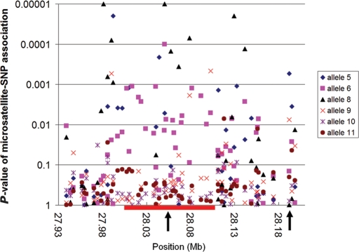
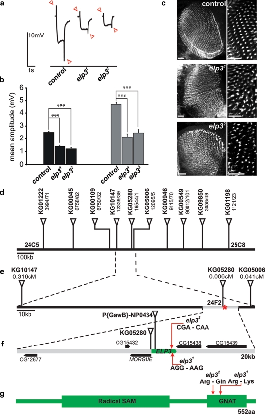
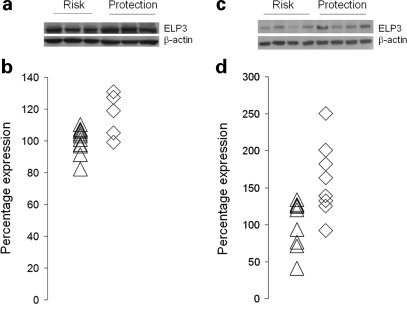
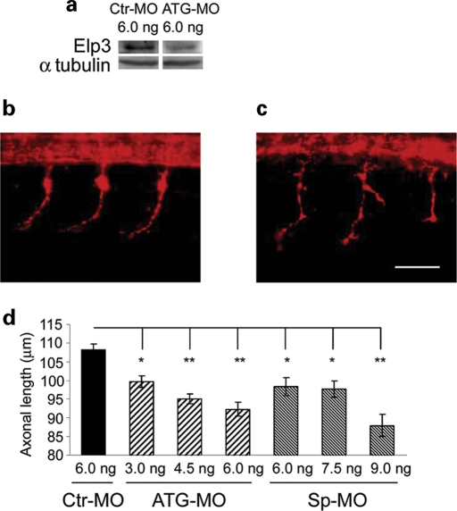
Amyotrophic lateral sclerosis (ALS) is a spontaneous, relentlessly progressive motor neuron disease, usually resulting in death from respiratory failure within 3 years. Variation in the genes SOD1 and TARDBP accounts for a small percentage of cases, and other genes have shown association in both candidate gene and genome-wide studies, but the genetic causes remain largely unknown. We have performed two independent parallel studies, both implicating the RNA polymerase II component, ELP3, in axonal biology and neuronal degeneration. In the first, an association study of 1884 microsatellite markers, allelic variants of ELP3 were associated with ALS in three human populations comprising 1483 people (P=1.96 x 10(-9)). In the second, an independent mutagenesis screen in Drosophila for genes important in neuronal communication and survival identified two different loss of function mutations, both in ELP3 (R475K and R456K). Furthermore, knock down of ELP3 protein levels using antisense morpholinos in zebrafish embryos resulted in dose-dependent motor axonal abnormalities [Pearson correlation: -0.49, P=1.83 x 10(-12) (start codon morpholino) and -0.46, P=4.05 x 10(-9) (splice-site morpholino), and in humans, risk-associated ELP3 genotypes correlated with reduced brain ELP3 expression (P=0.01). These findings add to the growing body of evidence implicating the RNA processing pathway in neurodegeneration and suggest a critical role for ELP3 in neuron biology and of ELP3 variants in ALS.


Figures

Figure 1.
Figure 1.
A scatterplot of the linkage disequilibrium between common alleles of microsatellite D8S1820 and neighboring SNPs in controls of the study population. The position of the ELP3 gene is shown as a red bar below the X-axis. Marker D8S1820 is marked by the central vertical arrow and rs12682496 by the right vertical arrow. Because of the multi-allelic nature of microsatellite markers it is difficult to show patterns of LD using the conventional triangle plots used for SNPs (but see Supplementary Material, Figs S5 and S6). This graph plots the pairwise LD between each SNP and the common microsatellite alleles, with the strength of LD represented by the P-value for a χ2 test of association. As can be seen, the pattern of LD with neighboring SNPs is complex, the strength of LD varies for different alleles, and LD may extend long distances.
Figure 2.
Figure 2.
Drosophila ELP3 mutants identified in a screen for defects in neuronal communication. (A and B) ERG recordings and quantification of ‘on’ and ‘off’ (arrowheads in A) amplitudes of control and ELP3 mutant eyes. Black: on- and grey off-transients. Controls n = 48, elp31 n = 59, elp32 n = 57. Error bars indicate standard error of the mean. t-test: Control-elp31, on: P = 1.86 × 10⁻¹⁶, off: P = 1.78 × 10⁻¹⁴; control-elp32, on: P = 1.58 × 10⁻¹⁹, off: P = 7.90 × 10⁻¹⁰. (C) Confocal microscopy showing the photoreceptor axon projections in the medulla labeled with anti-chaoptin. In the mutants photoreceptors arrive and synapse in the medulla, but the synapses are not properly organized in rows. Scale bars 20 µm. (D) Mapping of ELP3 mutations. KG P-elements used for fine-mapping located in the 24C5-8 cytological region. Numbers under markers are tested/recombinant flies. (E) Recombination distances (cM) for the three P-elements closest to the mutant phenotype (lethality). The position of ELP3 in relation to KG05280 is indicated with a red asterisk and the 20 kb sequenced region as a grey bar. (F) ELP3 mutations (arrows). No mutations were found in surrounding genes. (G) Schematic representation of fly ELP3 (552aa) and mutations elp31 and elp32.
Figure 3.
Figure 3.
Western blot analysis of ELP3 protein expression in human control cerebellar tissue and ALS motor cortex. (A) Western blot analysis of cerebellar tissue samples from controls carrying risk-associated alleles (Risk) or at least one protection-associated allele (Protection). (B) Expression of ELP3 protein as a ratio to β-actin in cerebellar tissue from controls carrying risk-associated alleles (triangles, n = 13) or at least one protection-associated allele (diamonds, n = 5). (C) Western blot analysis of ALS motor cortex tissue samples carrying risk-associated alleles (Risk) or at least one protection-associated allele (Protection). (D) Expression of ELP3 protein as a ratio to β-actin in ALS motor cortex samples carrying risk-associated alleles (triangles, n = 9) or at least one protection-associated allele (diamonds, n = 8).
Figure 4.
Figure 4.
Morpholino-induced knockdown of ELP3 affects motor neuron axonal branching and length. (A) Western blot of ELP3 following treatment with Ctr-MO and ATG-MO. Maximal ELP3 knockdown was 44%. (B) ELP3 knockdown by Sp-MO and ATG-MO resulted in increased branching of motor axons (right) compared with control (left). (C) There was a dose-dependent decrease in axonal length of motor neurons for both Sp-MO and ATG-MO. Results show SEM (*P < 0.01; **P < 1.0 × 10⁻⁷). P-values at each dose of ATG-MO compared with 6.0 ng Ctr-MO were 3.0 ng: P = 0.0024; 4.5 ng: P = 1.19 × 10⁻⁸; 6.0 ng: P = 7.68 × 10⁻¹¹. P-values at each dose of Sp-MO compared with 6.0 ng Ctr-MO were 6.0 ng: P = 0.0084; 7.5 ng: P = 0.0039; 9.0 ng, P = 6.94 × 10⁻⁹. Scale bar 50 µm.

References

    1. Simpson C.L., Al-Chalabi A. Amyotrophic lateral sclerosis as a complex genetic disease. Biochim. Biophys. Acta. 2006;1762:973–985. - PubMed
    1. Sreedharan J., Blair I.P., Tripathi V.B., Hu X., Vance C., Rogelj B., Ackerley S., Durnall J.C., Williams K.L., Buratti E., et al. TDP-43 mutations in familial and sporadic amyotrophic lateral sclerosis. Science. 2008;319:1668–1672. - PMC - PubMed
    1. Kabashi E., Valdmanis P.N., Dion P., Spiegelman D., McConkey B.J., Velde C.V., Bouchard J.P., Lacomblez L., Pochigaeva K., Salachas F., et al. TARDBP mutations in individuals with sporadic and familial amyotrophic lateral sclerosis. Nat. Genet. 2008;40:572–574. - PubMed
    1. Schymick J.C., Scholz S.W., Fung H.C., Britton A., Arepalli S., Gibbs J.R., Lombardo F., Matarin M., Kasperaviciute D., Hernandez D.G., et al. Genome-wide genotyping in amyotrophic lateral sclerosis and neurologically normal controls: first stage analysis and public release of data. Lancet Neurol. 2007;6:322–328. - PubMed
    1. Dunckley T., Huentelman M.J., Craig D.W., Pearson J.V., Szelinger S., Joshipura K., Halperin R.F., Stamper C., Jensen K.R., Letizia D., et al. Whole-genome analysis of sporadic amyotrophic lateral sclerosis. N. Engl. J. Med. 2007;357:775–788. - PubMed

Publication types

MeSH terms