

doi: 10.1128/EC.00267-08.
Epub 2008 Nov 7.
Apicoplast and mitochondrion in gametocytogenesis of Plasmodium falciparum
Affiliations
- PMID: 18996983
- PMCID: PMC2620748
- DOI: 10.1128/EC.00267-08
Item in Clipboard
Apicoplast and mitochondrion in gametocytogenesis of Plasmodium falciparum
Eukaryot Cell.
2009 Jan.
Abstract
Live cell imaging of human malaria parasites Plasmodium falciparum during gametocytogenesis revealed that the apicoplast does not grow, whereas the mitochondrion undergoes remarkable morphological development. A close connection of the two organelles is consistently maintained. The apicoplast and mitochondrion are not components of the male gametes, suggesting maternal inheritance.
Figures
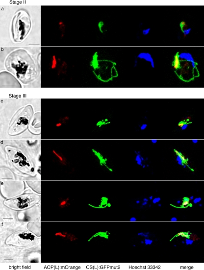
Live cell images of 3D7 ACP(L):mOrange-CS(L):GFPmut2 gametocytes at stage II (a and b) and stage III (c to f), showing a small apicoplast (red) that is always encased by the mitochondrion (green). At stage III, the apicoplast is round (c and e) or slightly elongated (d and f). In contrast, the mitochondrion forms branches from the cluster that encase the apicoplast. Sometimes the GFP labeling for the mitochondrion is heterologous, but continuous (c and d). Scale bars, 3 μm.
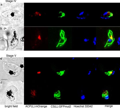
Live cell images of 3D7 ACP(L):mOrange-CS(L):GFPmut2, at stage IV (a and b) and stage V (c and d), showing the apicoplast (red) and mitochondrion (green) association in macrogametocytes (a and c) and microgametocytes (b and d). The apicoplast (red) does not elongate or branch but stays small. Often, the mOrange signal for the apicoplast is weak in the mature microgametocyte (d). Scale bars, 3 μm.
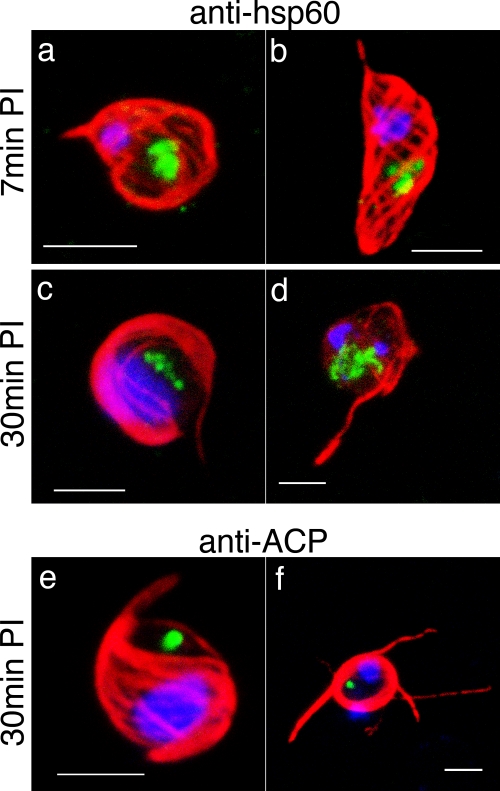
Exflagellated microgametes. Immunofluorescence images of the mitochondrion in NF54 parasites, fixed 7 min (a and b) or 30 min (c to f) after exflagellation induction. (a to d) The mitochondrion (labeled with the anti-Hsp60 antibody [green]) stays single even after each microgamete (whose axoneme is labeled with the anti-alpha tubulin antibody [red]) is formed (a and b) and exflagellated (c and d). (e and f) The apicoplast (labeled with the anti-ACP antibody [green]) stays single and does not enter the exflagellated microgametes (whose axoneme is labeled with the anti-alpha tubulin antibody [red]). Scale bars, 3 μm.
Similar articles
-
Does the shape of Plasmodium falciparum gametocytes have a function?Med Hypotheses. 2004;62(4):618-9. doi: 10.1016/j.mehy.2003.11.011. Med Hypotheses. 2004. PMID: 15050117
-
The dynamin-related protein PfDyn2 is essential for both apicoplast and mitochondrial fission in Plasmodium falciparum.mBio. 2025 Jan 8;16(1):e0303624. doi: 10.1128/mbio.03036-24. Epub 2024 Nov 29. mBio. 2025. PMID: 39611847 Free PMC article.
-
Malaria, Plasmodium falciparum and its apicoplast.Biochem Soc Trans. 2010 Jun;38(3):775-82. doi: 10.1042/BST0380775. Biochem Soc Trans. 2010. PMID: 20491664 Review.
-
In vivo profiles in malaria are consistent with a novel physiological state.Proc Natl Acad Sci U S A. 2009 Jul 7;106(27):E70; author reply E71-2. doi: 10.1073/pnas.0904478106. Epub 2009 Jul 1. Proc Natl Acad Sci U S A. 2009. PMID: 19570993 Free PMC article. No abstract available.
-
Plasmodium falciparum apicoplast drugs: targets or off-targets?Chem Rev. 2012 Mar 14;112(3):1269-83. doi: 10.1021/cr200258w. Epub 2011 Oct 25. Chem Rev. 2012. PMID: 22026508 Review. No abstract available.
Cited by
-
The evolution, metabolism and functions of the apicoplast.Philos Trans R Soc Lond B Biol Sci. 2010 Mar 12;365(1541):749-63. doi: 10.1098/rstb.2009.0273. Philos Trans R Soc Lond B Biol Sci. 2010. PMID: 20124342 Free PMC article. Review.
-
Identification of MMV malaria box inhibitors of plasmodium falciparum early-stage gametocytes using a luciferase-based high-throughput assay.Antimicrob Agents Chemother. 2013 Dec;57(12):6050-62. doi: 10.1128/AAC.00870-13. Epub 2013 Sep 23. Antimicrob Agents Chemother. 2013. PMID: 24060871 Free PMC article.
-
Plasmodium falciparum gametocytes display global chromatin remodelling during sexual differentiation.BMC Biol. 2023 Apr 3;21(1):65. doi: 10.1186/s12915-023-01568-4. BMC Biol. 2023. PMID: 37013531 Free PMC article.
-
A paternal lactate dehydrogenase critically enhances male gametogenesis and malaria transmission.Sci Rep. 2025 Jul 2;15(1):23283. doi: 10.1038/s41598-025-05832-1. Sci Rep. 2025. PMID: 40603939 Free PMC article.
-
Targeting Gametocytes of the Malaria Parasite Plasmodium falciparum in a Functional Genomics Era: Next Steps.Pathogens. 2021 Mar 16;10(3):346. doi: 10.3390/pathogens10030346. Pathogens. 2021. PMID: 33809464 Free PMC article. Review.
References
-
- Aikawa, M. 1966. The fine structure of the erythrocytic stages of three avian malarial parasites, Plasmodium fallax, P. lophurae, and P. cathemerium. Am. J. Trop. Med. Hyg. 15449-471. - PubMed
-
- Alano, P. 2007. Plasmodium falciparum gametocytes: still many secrets of a hidden life. Mol. Microbiol. 66291-302. - PubMed
-
- Bannister, L. H., J. M. Hopkins, R. E. Fowler, S. Krishna, and G. H. Mitchell. 2000. A brief illustrated guide to the ultrastructure of Plasmodium falciparum asexual blood stages. Parasitol. Today 16427-433. - PubMed
-
- Biagini, G. A., N. Fisher, N. Berry, P. A. Stocks, B. Meunier, D. P. Williams, R. Bonar-Law, P. G. Bray, A. Owen, P. M. O'Neill, and S. A. Ward. 2008. Acridinediones: selective and potent inhibitors of the malaria parasite mitochondrial bc1 complex. Mol. Pharmacol. 731347-1355. - PubMed
-
- Creasey, A., K. Mendis, J. Carlton, D. Williamson, I. Wilson, and R. Carter. 1994. Maternal inheritance of extrachromosomal DNA in malaria parasites. Mol. Biochem. Parasitol. 6595-98. - PubMed
Publication types
MeSH terms
Grants and funding
LinkOut - more resources
Full Text Sources
