

In vivo water state measurements in breast cancer using broadband diffuse optical spectroscopy
- PMID: 18997265
- PMCID: PMC2586905
- DOI: 10.1088/0031-9155/53/23/005
In vivo water state measurements in breast cancer using broadband diffuse optical spectroscopy
Abstract
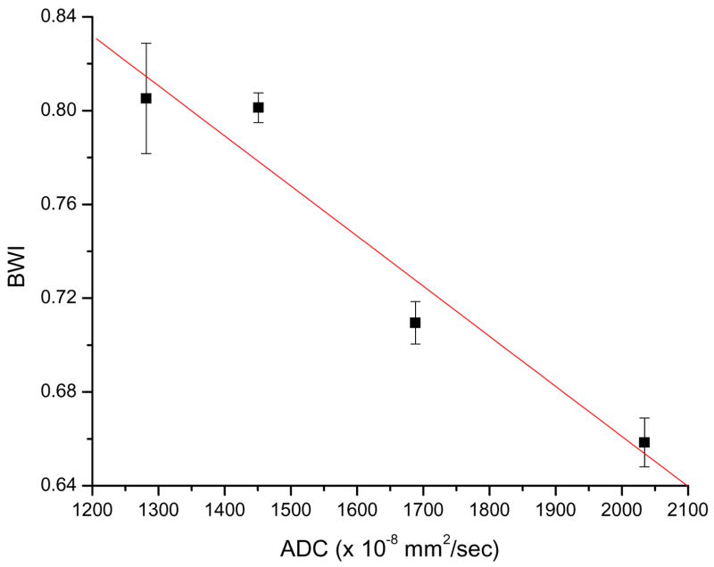
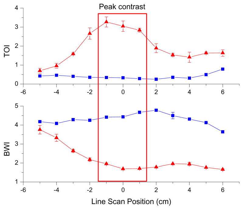
Structural changes in water molecules are related to physiological, anatomical and pathological properties of tissues. Near infrared (NIR) optical absorption methods are sensitive to water; however, detailed characterization of water in thick tissues is difficult to achieve because subtle spectral shifts can be obscured by multiple light scattering. In the NIR, a water absorption peak is observed around 975 nm. The precise NIR peak's shape and position are highly sensitive to water molecular disposition. We introduce a bound water index (BWI) that quantifies shifts observed in tissue water absorption spectra measured by broadband diffuse optical spectroscopy (DOS). DOS quantitatively measures light absorption and scattering spectra and therefore reveals bound water spectral shifts. BWI as a water state index was validated by comparing broadband DOS to magnetic resonance spectroscopy, diffusion-weighted MRI and conductivity in bound water tissue phantoms. Non-invasive DOS measurements of malignant and normal breast tissues performed in 18 subjects showed a significantly higher fraction of free water in malignant tissues (p < 0.0001) compared to normal tissues. BWI of breast cancer tissues inversely correlated with Nottingham-Bloom-Richardson histopathology scores. These results highlight broadband DOS sensitivity to molecular disposition of water and demonstrate the potential of BWI as a non-invasive in vivo index that correlates with tissue pathology.
Figures


Similar articles
-
Molecular imaging of water binding state and diffusion in breast cancer using diffuse optical spectroscopy and diffusion weighted MRI.J Biomed Opt. 2012 Jul;17(7):071304. doi: 10.1117/1.JBO.17.7.071304. J Biomed Opt. 2012. PMID: 22894465 Free PMC article.
-
Non-invasive tissue temperature measurements based on quantitative diffuse optical spectroscopy (DOS) of water.Phys Med Biol. 2010 Jul 7;55(13):3753-65. doi: 10.1088/0031-9155/55/13/012. Epub 2010 Jun 15. Phys Med Biol. 2010. PMID: 20551502 Free PMC article.
-
In vivo absorption, scattering, and physiologic properties of 58 malignant breast tumors determined by broadband diffuse optical spectroscopy.J Biomed Opt. 2006 Jul-Aug;11(4):044005. doi: 10.1117/1.2337546. J Biomed Opt. 2006. PMID: 16965162
-
Imaging in breast cancer: diffuse optics in breast cancer: detecting tumors in pre-menopausal women and monitoring neoadjuvant chemotherapy.Breast Cancer Res. 2005;7(6):279-85. doi: 10.1186/bcr1358. Epub 2005 Nov 28. Breast Cancer Res. 2005. PMID: 16457705 Free PMC article. Review.
-
The role of diffuse optical spectroscopy in the clinical management of breast cancer.Dis Markers. 2003-2004;19(2-3):95-105. doi: 10.1155/2004/460797. Dis Markers. 2003. PMID: 15096707 Free PMC article. Review.
Cited by
-
Breast composition measurement with a cadmium-zinc-telluride based spectral computed tomography system.Med Phys. 2012 Mar;39(3):1289-97. doi: 10.1118/1.3681273. Med Phys. 2012. PMID: 22380361 Free PMC article.
-
Frequency domain diffuse optical spectroscopy with a near-infrared tunable vertical cavity surface emitting laser.Opt Express. 2018 Aug 6;26(16):21033-21043. doi: 10.1364/OE.26.021033. Opt Express. 2018. PMID: 30119409 Free PMC article.
-
Macroscopic optical physiological parameters correlate with microscopic proliferation and vessel area breast cancer signatures.Breast Cancer Res. 2015 May 27;17:72. doi: 10.1186/s13058-015-0578-z. Breast Cancer Res. 2015. PMID: 26013572 Free PMC article.
-
Differential diagnosis of breast masses in South Korean premenopausal women using diffuse optical spectroscopic imaging.J Biomed Opt. 2016 Jul 1;21(7):74001. doi: 10.1117/1.JBO.21.7.074001. J Biomed Opt. 2016. PMID: 27436049 Free PMC article.
-
Tight-frame based iterative image reconstruction for spectral breast CT.Med Phys. 2013 Mar;40(3):031905. doi: 10.1118/1.4790468. Med Phys. 2013. PMID: 23464320 Free PMC article.
References
-
- Assaf Y, BeitYannai E, Shohami E, Berman E, Cohen Y. Diffusion- and T-2-weighted MRI of closed-head injury in rats: A time course study and correlation with histology. Magnetic Resonance Imaging. 1997;15:77–85. - PubMed
-
- Baumgartner G, et al. The impact of extracellular matrix on the chemoresistance of solid tumors — experimental and clinical results of hyaluronidase as additive to cytostatic chemotherapy. Cancer Lett. 1998;131:85–99. - PubMed
-
- Bellamy LJ. Advances in Infrared Group Frequencies. London: Chapman and Hall; 1968.
Publication types
MeSH terms
Grants and funding
LinkOut - more resources
Full Text Sources
Medical
Miscellaneous