

Kindlins: essential regulators of integrin signalling and cell-matrix adhesion
- PMID: 18997731
- PMCID: PMC2603460
- DOI: 10.1038/embor.2008.202
Kindlins: essential regulators of integrin signalling and cell-matrix adhesion
Abstract
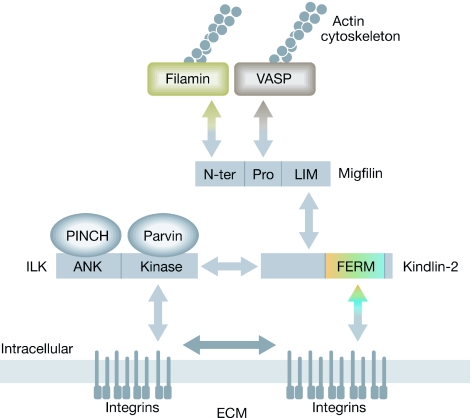
Integrin-mediated cell-ECM (extracellular matrix) adhesion is a fundamental process that controls cell behaviour. For correct cell-ECM adhesion, both the ligand-binding affinity and the spatial organization of integrins must be precisely controlled; how integrins are regulated, however, is not completely understood. Kindlins constitute a family of evolutionarily conserved cytoplasmic components of cell-ECM adhesions that bind to beta-integrin cytoplasmic tails directly and cooperate with talin in integrin activation. In addition, kindlins interact with many components of cell-ECM adhesions--such as migfilin and integrin-linked kinase--to promote cytoskeletal reorganization. Loss of kindlins causes severe defects in integrin signalling, cell-ECM adhesion and cytoskeletal organization, resulting in early embryonic lethality (kindlin-2), postnatal lethality (kindlin-3) and Kindler syndrome (kindlin-1). It is therefore clear that kindlins, together with several other integrin-proximal proteins, are essential for integrin signalling and cell-ECM adhesion regulation.
Figures


References
-
- Calderwood DA, Zent R, Grant R, Rees DJ, Hynes RO, Ginsberg MH (1999) The Talin head domain binds to integrin beta subunit cytoplasmic tails and regulates integrin activation. J Biol Chem 274: 28071–28074 - PubMed
-
- Calderwood DA, Yan B, de Pereda JM, Alvarez BG, Fujioka Y, Liddington RC, Ginsberg MH (2002) The phosphotyrosine binding-like domain of talin activates integrins. J Biol Chem 277: 21749–21758 - PubMed
-
- Chen Y, Djaffar I, Pidard D, Steiner B, Cieutat A, Caen JP, Rosa J (1992) Ser-752→Pro mutation in the cytoplasmic domain of integrin beta 3 subunit and defective activation of platelet integrin alpha IIb beta 3 (glycoprotein IIb-IIIa) in a variant of Glanzmann thrombasthenia. Proc Natl Acad Sci USA 89: 10169–10173 - PMC - PubMed
-
- Chen YP, O'Toole TE, Ylanne J, Rosa JP, Ginsberg MH (1994) A point mutation in the integrin beta 3 cytoplasmic domain (S752→P) impairs bidirectional signaling through alpha IIb beta 3 (platelet glycoprotein IIb-IIIa). Blood 84: 1857–1865 - PubMed
-
- Critchley DR (2005) Genetic, biochemical and structural approaches to talin function. Biochem Soc Trans 33: 1308–1312 - PubMed
Publication types
MeSH terms
Substances
Grants and funding
LinkOut - more resources
Full Text Sources
Other Literature Sources
