. 2008 Dec 11;456(7223):814-8.
doi: 10.1038/nature07445. Epub 2008 Nov 9.

Deletion of vascular endothelial growth factor in myeloid cells accelerates tumorigenesis

Affiliations

Deletion of vascular endothelial growth factor in myeloid cells accelerates tumorigenesis

Christian Stockmann et al. Nature. .

Abstract

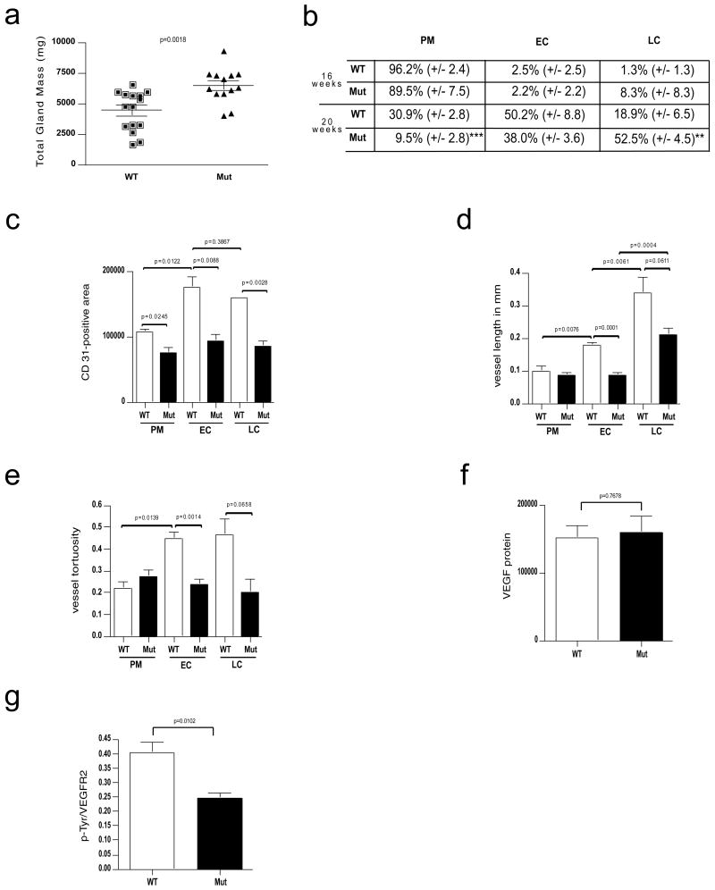
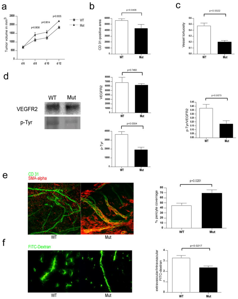
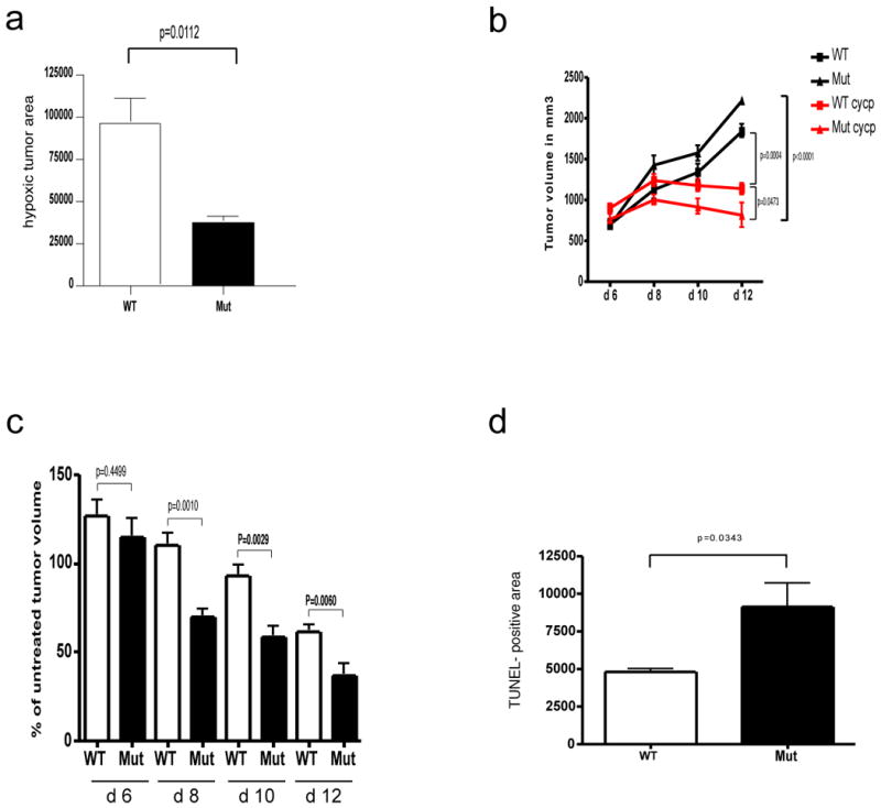
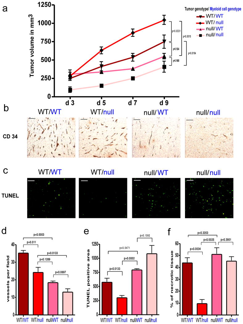
Angiogenesis and the development of a vascular network are required for tumour progression, and they involve the release of angiogenic factors, including vascular endothelial growth factor (VEGF-A), from both malignant and stromal cell types. Infiltration by cells of the myeloid lineage is a hallmark of many tumours, and in many cases the macrophages in these infiltrates express VEGF-A. Here we show that the deletion of inflammatory-cell-derived VEGF-A attenuates the formation of a typical high-density vessel network, thus blocking the angiogenic switch in solid tumours in mice. Vasculature in tumours lacking myeloid-cell-derived VEGF-A was less tortuous, with increased pericyte coverage and decreased vessel length, indicating vascular normalization. In addition, loss of myeloid-derived VEGF-A decreases the phosphorylation of VEGF receptor 2 (VEGFR2) in tumours, even though overall VEGF-A levels in the tumours are unaffected. However, deletion of myeloid-cell VEGF-A resulted in an accelerated tumour progression in multiple subcutaneous isograft models and an autochthonous transgenic model of mammary tumorigenesis, with less overall tumour cell death and decreased tumour hypoxia. Furthermore, loss of myeloid-cell VEGF-A increased the susceptibility of tumours to chemotherapeutic cytotoxicity. This shows that myeloid-derived VEGF-A is essential for the tumorigenic alteration of vasculature and signalling to VEGFR2, and that these changes act to retard, not promote, tumour progression.


Figures

Figure 1
Figure 1
Deletion of VEGF in myeloid cells results in reduced vascularization but accelerated progression of mammary tumors. a, Total tumor mass of PyMT-WT mice (n=15) and PyMT-Mut mice (n=13) at the age of 20 weeks. b, Distribution of PyMT mammary tumors at prototypical premalignant (PM) lesions, malignant early carcinoma (EC) and late carcinoma (LC) stages in percent (±s.e.m.) between genotypes and at the age of 16 (n=4) and 20 weeks (PyMT-WT n=4, PyMT-Mut n=9). c, Quantitative analysis of the area covered by CD 31-positive cells within a tumor section for each stage (n=4). d, Development of vessel length in PyMT-tumors during malignant progression as determined by tracing CD 31-positive vessels that were exposed in a longitudinal cut (n=4). e, Analysis of vessel tortuosity based on CD 31-stained tumor sections (n=4). f, Quantitative analysis of the VEGF signal (PyMT-WT n=7, PyMT-Mut n=4). g, ratio of p-Tyr and VEGFR2 signal intensities as a measure of receptor activation (PyMT-WT n=7, PyMT-Mut n=4). Scale bars, 100 μm; **p<0.01, ***p<0.001; error bars, s.e.m.
Figure 2
Figure 2
Deletion of VEGF in myeloid cells leads to a normalized vasculature and higher tumor volumes in Lewis Lung Carcinoma isografts. a, Growth curve analysis of Lewis Lung Carcinoma (LLC) tumors injected subcutaneously in WT and Mut mice (n>7 for each group). b, Quantitative analysis of CD 31-positive endothelial cells (n=4). c, Determination of blood vessel tortuosity in LLC tumors. d, Left: Immunoblotting for VEGFR2 and phosphotyrosine (p-Tyr) after Immunoprecipitation of VEGFR2 from LLC tumor lysates. Center: Quantitative analysis of VEGFR2 (upper) and phosphotyrosine (lower) signals. Right: Ratio of p-Tyr and VEGFR2 signal intensities as a measure of receptor activation (WT n=8, Mut n=7). e, Left: Confocal microscopy images of simultaneous immunodetection of endothelial cells and pericytes in LLC tumors with the specific markers CD 31 and smooth muscle actin-alpha (SMA-alpha). Right: Pericyte coverage as assessed by SMA-alpha/CD 31 co-localization (n=4). f, Left: Fluorescent microscopy images of a FITC-dextran angiography on LLC isografts. Right: Ratio of extravascular over intravascular FITC-dextran as a measure of vascular permeability (WT n=6, Mut n=4). Scale bar, 100 μm; error bars, s.e.m.
Figure 3
Figure 3
Deletion of VEGF myeloid cells results in reduced hypoxia and increased susceptibility of LLC tumors to cytotoxic agents. a, Quantitative analysis of hypoxic tumor areas (WT n=6, Mut n=4). b, Growth curve analysis of LLC isografts from WT and Mut animals (n>4 for each group) treated with Cyclophosphamide (CYCP) (170mg/kg) at day 6, 8 and 10 after tumor implantation. c, Response of tumors from WT and Mut mice to CYCP treatment expressed as percentage of treated tumor volume to untreated tumor volume at specified time points. d, Quantitative analysis of TUNEL positive on CYCP-treated LLC isografts (n=4). Scale bar, 100 μm; error bars, s.e.m.
Figure 4
Figure 4
Effect of tumor cell-derived versus myeloid cell-derived VEGF on tumor angiogenesis and growth. a, Growth curve analysis of wildtype (WT) and VEGF nullizygous (null) fibrosarcoma isografts (genotype labeled in black) implanted into WT-mice or Mut-mice (null) with a myeloid cell-specific deletion of VEGF (genotype labeled in blue) (n=4 for each group). b, CD 34 immunostaining on fibrosarcoma isografts. c, Detection of apoptotic cells in fibrosarcomas by TUNEL-staining. d, Quantitative analysis of CD 34-positive blood vessels (n=4). e, Quantification of TUNEL-positive cells (n=4). f, Assessment of tumor necrosis on fibrosarcoma midline sections. Shown is the perimeter of necrotic areas expressed as percentage of total tumor perimeter (n=4). Scale bar, 100 μm; error bars, s.e.m.

Similar articles

Cited by

References

    1. Folkman J. Angiogenesis. Annu Rev Med. 2006;57:1–18. - PubMed
    1. Lewis JS, Landers RJ, Underwood JC, Harris AL, Lewis CE. Expression of vascular endothelial growth factor by macrophages is up-regulated in poorly vascularized areas of breast carcinomas. J Pathol. 2000;192(2):150–158. - PubMed
    1. Gerber H, et al. VEGF couples hypertrophic cartilage remodeling, ossification and angiogenesis during endochondral bone formation. Nature Medicine. 1999;5:623–628. - PubMed
    1. Clausen BE, Burkhardt C, Reith W, Renkawitz R, Forster I. Conditional gene targeting in macrophages and granulocytes using LysMcre mice. Transgenic Res. 1999;8(4):265–277. - PubMed
    1. Cramer T, et al. HIF-1alpha is essential for myeloid cell-mediated inflammation. Cell. 2003;112(5):645–657. - PMC - PubMed

Publication types

MeSH terms

Substances