. 2008 Dec;14(12):1390-5.
doi: 10.1038/nm.1779. Epub 2008 Nov 9.

Control of HIV-1 immune escape by CD8 T cells expressing enhanced T-cell receptor

Affiliations

Control of HIV-1 immune escape by CD8 T cells expressing enhanced T-cell receptor

Angel Varela-Rohena et al. Nat Med. 2008 Dec.

Abstract

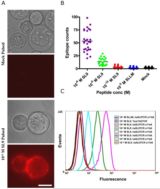
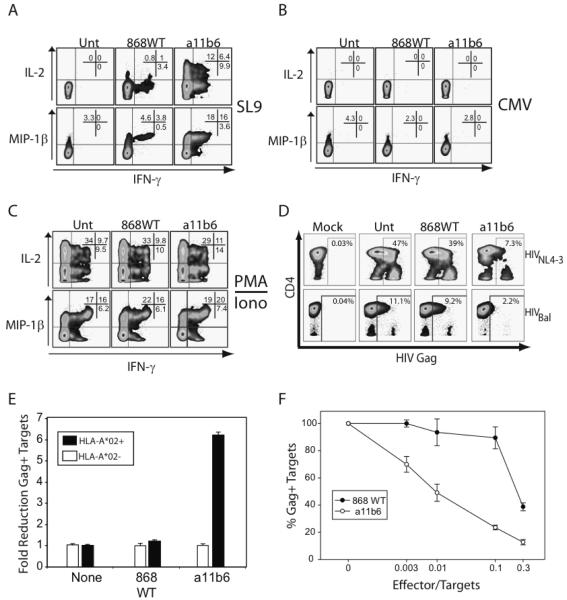
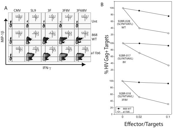
HIV's considerable capacity to vary its HLA-I-restricted peptide antigens allows it to escape from host cytotoxic T lymphocytes (CTLs). Nevertheless, therapeutics able to target HLA-I-associated antigens, with specificity for the spectrum of preferred CTL escape mutants, could prove effective. Here we use phage display to isolate and enhance a T-cell antigen receptor (TCR) originating from a CTL line derived from an infected person and specific for the immunodominant HLA-A(*)02-restricted, HIVgag-specific peptide SLYNTVATL (SL9). High-affinity (K(D) < 400 pM) TCRs were produced that bound with a half-life in excess of 2.5 h, retained specificity, targeted HIV-infected cells and recognized all common escape variants of this epitope. CD8 T cells transduced with this supraphysiologic TCR produced a greater range of soluble factors and more interleukin-2 than those transduced with natural SL9-specific TCR, and they effectively controlled wild-type and mutant strains of HIV at effector-to-target ratios that could be achieved by T-cell therapy.


Figures

Figure 1
Figure 1. SL9 specific cell staining with high affinity TCR
A. Mock-pulsed (top two panels) and SL9-pulsed (bottom two panels) T2 cells were stained with haSL9TCR a11b6-bio/streptavidin-PE and visualized by brightfield (panels 1&3) or fluorescence (panels 2&4). The entire 3-dimensional surface of the cell was imaged by fluorescence microscopy. The fluorescent image in panels 2&4 is a representation of a single plane through the cell while the brightfield image shows the location of the cells. The several hundred PE molecules in the single plane shown in panel 4 make exact counting difficult for this level of antigen. We estimate that this pulsing with 10−6M SL9 peptide loads each cell with >5000 SL9 epitopes. Bar =10 μM. B. Total antigen levels on SL9-pulsed T2 cells as determined by 3D fluorescence microscopy through 20 Z-planes. Cells were pulsed with indicated concentrations of SLYNTVATL and SLLMWITQV (SLLM) or mock pulsed and then stained with haSL9TCR a11b6-bio/streptavidin-PE as described in the materials and methods. C. FACS analysis of haSL9TCR a11b6-bio staining of T2 cells pulsed with a titration of SL9 peptide. Control stains (SLLMWITQV peptide + haSL9TCR a11b6 TCR-bio, or SL9 peptide + Tax(134) TCR-bio; are shown to indicate background staining.
Figure 2
Figure 2. CD8 T-cells expressing supraphysiologic SL9-specific TCRs show an increased frequency of polyfunctional cells and an increased ability to control HIV spread
A, B, C. CD8+ T-cells were transduced with indicated TCR and then stimulated by K562 cells expressing HLA-A*0201 (KT.A2) loaded with 50 pg/ml SLYNTVATL (SL9) (A) or CMV (NLVPMVATV) (B) for 5 hours and expression of IFN-γ, IL-2 and MIP-1β was measured by intracellular cytokine staining. PMA + ionomycin (C) stimulation for 5 hours was used as a positive control D. Untransduced or SL9-specific TCR-transduced CD8 T-cells were added as effectors to a culture of HIVNL4-3 (TOP) or HIVBal (BOTTOM) infected HLA-A*02+ CD4 T-cell targets at a ratio of 1:20 (E:T). Eight days after co-culturing, HIV infection was assessed by intracellular stain for HIVgag. Data is representative of three independent experiments E. HLA-A*02+ and HLA-A*02- targets were infected and the indicated SL9-specific TCR transduced effectors were added at a 1:20 E:T ratio. The fold reduction of HIVgag+ cells was calculated by dividing the number of HIVgag+ cells in cultures mixed with untransduced CD8 T-cells by the number of HIVgag+ cells in the other indicated cultures. Error bars represent the standard error of one experiment performed in triplicate, and the data is representative of 2 independent experiments. F. HIV infection was established in an HLA-A*02+ CD4 T-cell culture and TCR transduced effectors were added at the ratios shown. The percentage of HIVgag+ cells was compared between the conditions and standardized to the addition of untransduced effectors. Error bars represent standard error of triplicate measurements and the graph represent one of three independent experiments.
Figure 3
Figure 3. High avidity TCR-transduced CD8 T-cells can effectively recognize SL9 escape mutant peptides and suppress infection by SL9 escape HIV-1 variants
A. KT.A2 targets were loaded with a control peptide (CMV pp65), SLYNTVATL (SL9) or naturally occurring escape mutant peptides: SLFNTVATL (3F), SLFNTVAVL (3F8V), and SLFNTIAVL (3F6I8V). Effector CD8 T-cells were added and cytokine production after 5 hour stimulation is shown. B. HIV infection was established in PHA blasted HLA-A*02+ CD4 T-cells with three primary isolates with mutated SL9 epitopes. The indicated TCR-transduced CD8 T-cells were added to a final target to effector ratio of 10:1 or 50:1. The percentage of HIVgag+ cells was compared between the conditions and standardized to the addition of untransduced effectors. Data is representative of at least two independent experiments.

References

    1. Goulder PJ, Watkins DI. HIV and SIV CTL escape: implications for vaccine design. Nat Rev Immunol. 2004;4:630–40. - PubMed
    1. Sewell AK, Price DA, Oxenius A, Kelleher AD, Phillips RE. Cytotoxic T lymphocyte responses to human immunodeficiency virus: control and escape. Stem Cells. 2000;18:230–44. - PubMed
    1. Phillips RE, et al. Human immunodeficiency virus genetic variation that can escape cytotoxic T cell recognition. Nature. 1991;354:453–9. - PubMed
    1. Price DA, et al. The influence of antigenic variation on cytotoxic T lymphocyte responses in HIV-1 infection. J Mol Med. 1998;76:699–708. - PubMed
    1. Cohen GB, et al. The selective downregulation of class I major histocompatibility complex proteins by HIV-1 protects HIV-infected cells from NK cells. Immunity. 1999;10:661–71. - PubMed

Publication types