. 2008 Dec;15(12):1278-86.
doi: 10.1038/nsmb.1512. Epub 2008 Nov 9.

Structural basis for midbody targeting of spastin by the ESCRT-III protein CHMP1B

Affiliations

Structural basis for midbody targeting of spastin by the ESCRT-III protein CHMP1B

Dong Yang et al. Nat Struct Mol Biol. 2008 Dec.

Abstract

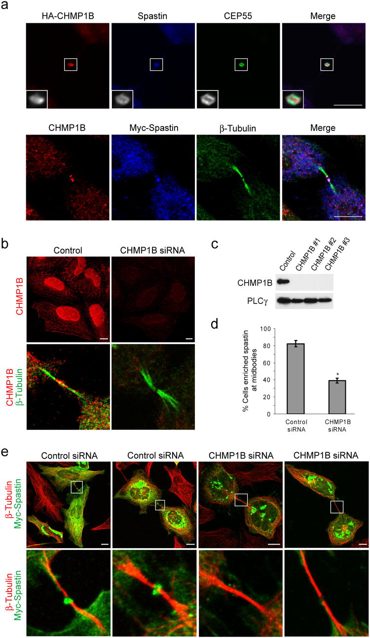
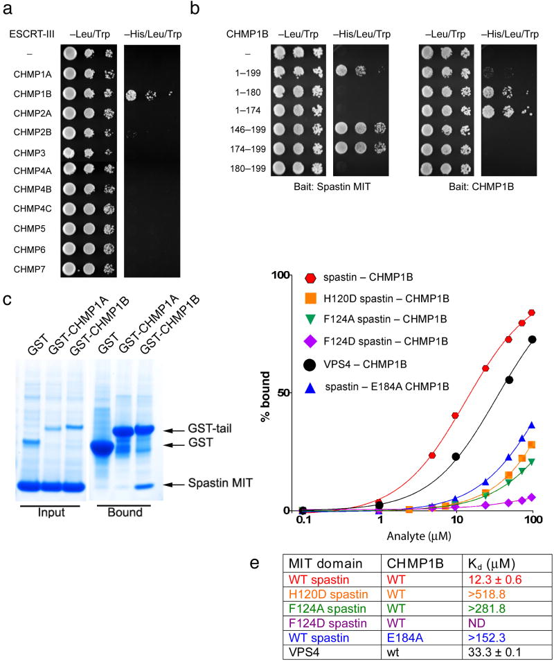
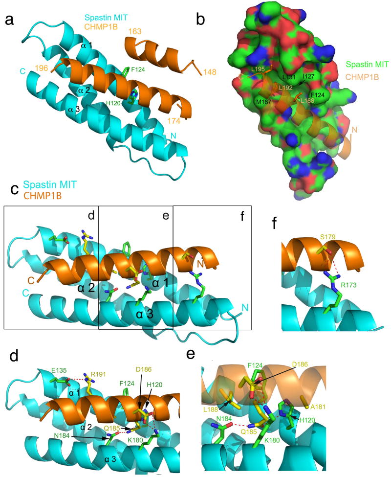
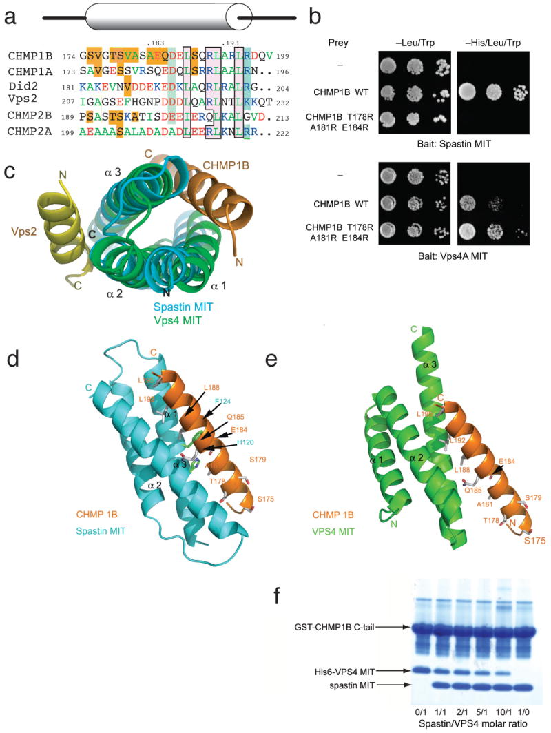
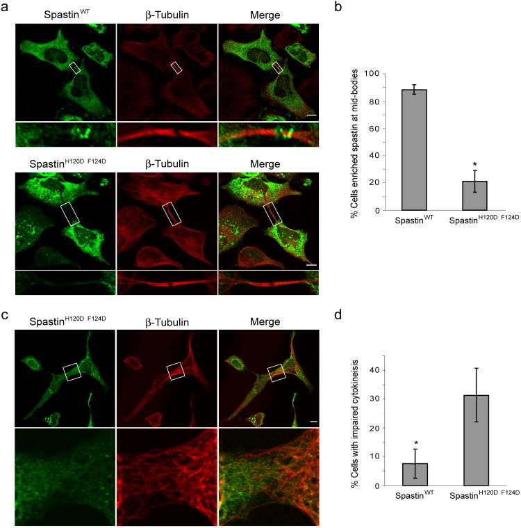
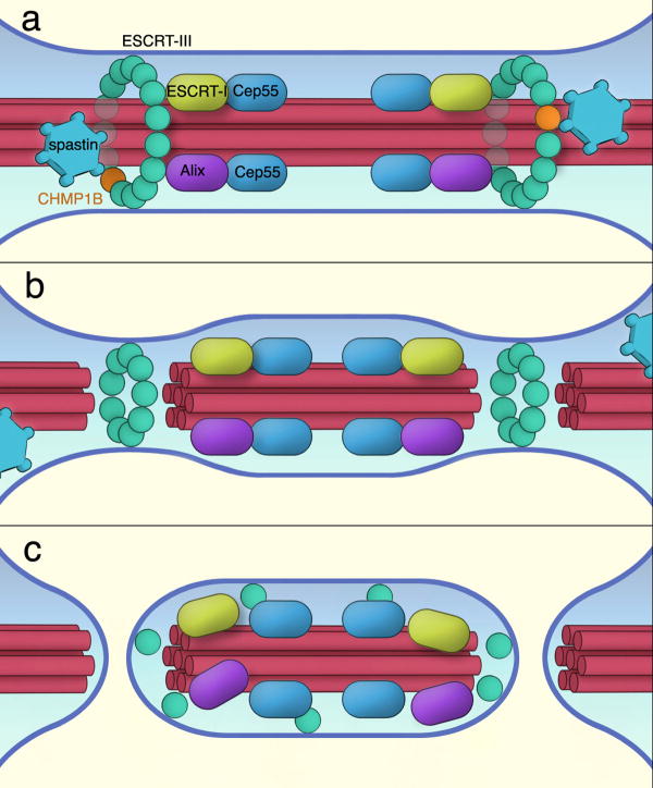
The endosomal sorting complex required for transport (ESCRT) machinery, including ESCRT-III, localizes to the midbody and participates in the membrane-abscission step of cytokinesis. The ESCRT-III protein charged multivesicular body protein 1B (CHMP1B) is required for recruitment of the MIT domain-containing protein spastin, a microtubule-severing enzyme, to the midbody. The 2.5-A structure of the C-terminal tail of CHMP1B with the MIT domain of spastin reveals a specific, high-affinity complex involving a noncanonical binding site between the first and third helices of the MIT domain. The structural interface is twice as large as that of the MIT domain of the VPS4-CHMP complex, consistent with the high affinity of the interaction. A series of unique hydrogen-bonding interactions and close packing of small side chains discriminate against the other ten human ESCRT-III subunits. Point mutants in the CHMP1B binding site of spastin block recruitment of spastin to the midbody and impair cytokinesis.


Figures

Figure 1
Figure 1. CHMP1B is present at midbodies and colocalizes with and recruits spastin
(a) Top panels, HeLa cells expressing HA-CHMP1B were immunostained for HA-epitope (red) as well as endogenous spastin (blue) and CEP55 (green). Lower panels, Cells expressing Myc-Spastin were immunostained for endogenous CHMP1B (red), Myc-epitope (blue), and β-tubulin (green). Bar, 10 μm. (b) Lysates from HeLa cells transfected with either of three different siRNAs specific for CHMP1B or else control siRNA were immunoblotted for CHMP1B. Equal protein loading was monitored by immunoblotting for PLCγ. (c) HeLa cells transfected with control siRNA or CHMP1B siRNA #3 were immunostained with anti-CHMP1B antibodies (red; top panels). Enrichment of CHMP1B at midbodies, as revealed by β-tubulin co-staining (green; bottom panels), is substantially decreased in CHMP1B-depleted cells (red; bottom panels). Bar, 10 μm. (d and e) HeLa cells transfected with either control or CHMP1B siRNA were re-transfected with Myc-spastin. (d) Quantification of spastin enrichment at midbodies in CHMP1B or control siRNA treated cells (n=3; 100 cells per experiment; ±SD). *p<0.001. (e) Midbodies, as identified by β-tubulin staining (red), show a lack of spastin (green) enrichment in CHMP1B-depleted cells (right two panels) as compared with control cells (left two panels). The boxed area is enlarged in the lower panels. Bar, 10 μm.
Figure 2
Figure 2. Spastin-CHMP1B interactions
(a) Yeast two-hybrid interactions between the spastin MIT domain bait and the indicated CHMP prey constructs were assayed using the HIS3 reporter (sequential 10-fold yeast dilutions shown). (b) Spastin MIT and CHMP1B baits were tested for yeast two-hybrid interactions as in (a) with the indicted CHMP1B prey constructs. (c) GST-fusion of the CHMP1B-CTR, but not CHMP1A-CTR, pulls down the spastin MIT domain in vitro. (d) SPR analysis of spastin and VPS4A MIT domain binding to immobilized CHMP1B-CTR. The analyte is either the spastin or VPS4A MIT domain, as indicated. (e) Tabulated binding constants (Kd), or their lower limits.
Figure 3
Figure 3. Structure of the spastin MIT domain-CHMP1B complex
(a) Overall structure of the MIT domain (blue) and CHMP1B (orange). (b) Key MIM Leu residues bind to hydrophobic pockets in the groove between α1 and α3 of the MIT domain. The spastin MIT domain surface is colored green for carbon atoms, red for oxygen, and blue for nitrogen. Contiguous green regions indicated hydrophobic surfaces. (c) Overview of polar interactions between CHMP1B-CTR and spastin MIT domain. (d-f) Details of interactions as seen in insets (d-f) within panel (c).
Figure 4
Figure 4. Overlapping specificity determinants in MIT domain recognition
(a) Structure-based sequence alignment of CHMP1B and other MIM-containing ESCRT-III proteins. Residues are boxed in gold for spastin MIT-specific contacts, green for VPS4 MIT-specific contacts, and pink with black outlines for residues that make contacts with both MIT domains. Residue abbreviation typeface is colored according to residues properties: green, hydrophobic; red, negatively charged; and blue, positively charged. (b) Yeast two-hybrid analysis using the HIS3 reporter (10-fold yeast dilutions shown) investigating the indicated CHMP1B triple mutant that abolishes spastin MIT binding, but not VPS4A MIT binding. (c) The spastin MIT (blue)-CHMP1B (orange) complex is overlaid on the yeast Vps4 MIT (green)-Vps2 (yellow) complex using a structural alignment between the MIT domains to show the CHMP1B and Vps2 bind to different sites on opposite faces of the MIT domain. (d) and (e) show a side-by-side view of the spastin MIT-CHMP1B complex (d) and a docked model of the VPS4 MIT-CHMP1B complex (e), colored as in (c). (f) The spastin MIT domain competes with the VPS4 MIT domain for binding to GST- CHMP1B-CTR in vitro. Molar ratios of these two MIT domains are shown at the bottom of the figure. The concentration of the VPS4 MIT domain was held constant while the concentration of the spastin MIT domain was varied.
Figure 5
Figure 5. Spastin MIT domain mutant protein shows decreased enrichment at midbodies and alters cytokinesis
(a) HeLa cells expressing wild-type (WT) Myc-spastin (green; top panels) or else Myc-spastinH120D/F124D (green; bottom panels) were co-immunostained for Myc-epitope and β-tubulin. Myc-spastinH120D/F124D shows decreased enrichment at midbodies, as identified by β-tubulin. Boxed areas are enlarged in the lower panels. Bar, 10 μm. (b) Quantification of enrichment of wild-type versus MIT mutant spastin expressing cells (n=3; 100 cells per experiment; ±SD). *p=0.001. (c) HeLa cells expressing Myc-spastinH120D/F124D (green) exhibited impaired cytokinesis, with microtubules (red) often maintaining a connection between cells. The boxed area is enlarged in the lower panels. Bar, 10 μm. (d) Quantification of cytokinesis impairment, as defined by the persistence of cellular interconnections, in wild-type Myc-spastin versus Myc-spastinH120D/F124D expressing cells (n=3; 100 cells per experiment; ±SD). *p<0.05.
Figure 6
Figure 6. Speculative model for the role of the spastin-ESCRT-III interaction in cytokinesis
(a) The assembly of CEP55, ESCRT-I, and Alix at the midbody is postulated to recruit ESCRT-III (green spheres indicate generic ESCRT-III subunits). Co-assembly of CHMP1B (orange sphere) into the ESCRT-III array leads to allosteric activation of CHMP1B, exposing its C-terminal helix, and thereby to the recruitment of spastin (blue hexagon). The circular array of ESCRT-III proteins is depicted schematically as inspired by observations of their formation of spiral or helical arrays ,. Two CEP55-ESCRT-spastin assemblies are shown in accord with the double cluster visible in Fig. 1C and ,,. (b) Cleavage of the microtubules (red tubes) by spastin opens a path for ESCRT-III to sever the membrane neck. Direct proof that ESCRT-III severs membrane necks is lacking, but it is widely believed to have such activity, perhaps in conjunction with other ESCRT components . (c) Midbody resolution can occur on both sides leaving behind a free midbody fragment that is sometimes observed in cell culture, or it may occur stochastically on one side or the other.

References

    1. Saksena S, Sun J, Chu T, Emr SD. ESCRTing proteins in the endocytic pathway. Trends Biochem Sci. 2007;32:561–573. - PubMed
    1. Hurley JH. ESCRT Complexes and the Biogenesis of Multivesicular Bodies. Curr Opin Cell Biol. 2008;20:4–11. - PMC - PubMed
    1. Morita E, Sundquist WI. Retrovirus budding. Ann Rev Cell Dev Biol. 2004;20:395–425. - PubMed
    1. Carlton JG, Martin-Serrano J. Parallels between cytokinesis and retroviral budding: a role for the ESCRT machinery. Science. 2007;316:1908–1912. - PubMed
    1. Morita E, et al. Human ESCRT and ALIX proteins interact with proteins of the midbody and function in cytokinesis. EMBO J. 2007;26:4215–4227. - PMC - PubMed

Publication types