

doi: 10.1038/nmeth.1269.
Epub 2008 Nov 9.
Intravital imaging of metastatic behavior through a mammary imaging window
Affiliations
- PMID: 18997781
- PMCID: PMC2820719
- DOI: 10.1038/nmeth.1269
Item in Clipboard
Intravital imaging of metastatic behavior through a mammary imaging window
Nat Methods.
2008 Dec.
Abstract
We report a technique to evaluate the same tumor microenvironment over multiple intravital imaging sessions in living mice. We optically marked individual tumor cells expressing photoswitchable proteins in an orthotopic mammary carcinoma and followed them for extended periods through a mammary imaging window. We found that two distinct microenvironments in the same orthotopic mammary tumor affected differently the invasion and intravasation of tumor cells.
Figures
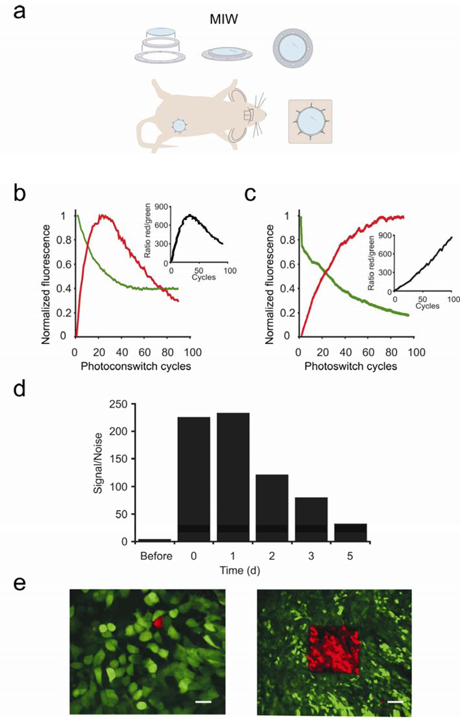
The Mammary Imaging Window (MIW) allows for long-term, high resolution imaging of the orthotopic tumors. (a) Components and the assembly of the MIW: a coverslip is mounted on a plastic frame consisting of two plastic rings and surgically implanted on top of the mammary gland or mammary tumor. (b,c) Average increase in red and decrease in green signal for Dendra2, as measured in a region of interest, in cells in vitro (b) or in vivo (c) upon photoswitching. The values were normalized to the highest fluorescent level in red and the initial fluorescent level in green. The insets show the ratio of the non-normalized red and green fluorescence. (d) Cells within Dendra2-tumors were photoswitched through the MIW and the red fluorescence was quantified before, immediately after (0 days) photoswitching and for the 5 subsequent days. The values were normalized to the red fluorescence level before photoswitching. (e) Photoswitching of Dendra2-expressing MTLn3 tumor cells in vivo can be done in regions of interest ranging from one cell (left panel, scale bar 10 µm) to hundreds of cells (right panel, scale bar 75 µm) through the MIW. Shown are combined images from the green and red channels using an OR-function: Only the pixels in the red channel that are above background are shown, and for all other pixels, the green channel is shown.
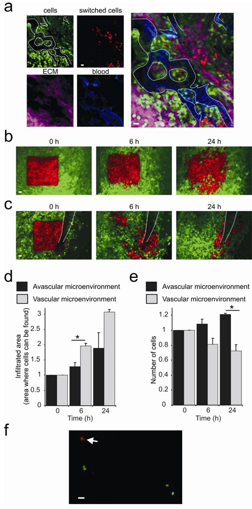
Photoswitching through the MIW is a tool for studying orthotopic tumor microenvironments. (a) By using Dendra2 as the label for tumor cells (red and green), Texas Red dextran for blood vessels (blue) and reflectance (purple) for extracellular matrix (ECM), one can define vascular microenvironments and monitor chosen cells inside them. (b,c) Non-photoswitched cells (green) and photoswitched cells (red) are shown at 0h, 6h and 24h after the photoswitch in avascular (b) and vascular (c) microenvironments (visible vessel indicated by white dotted lines). Shown are combined images from the green and red channels using an OR-function: Only the pixels in the red channel that are above background are shown, and for all other pixels, the green channel is shown. Scale bars 30 µm. The relative infiltration areas (d) and numbers (e) of photoswitched cells over time in vascular and avascular microenvironments. Error bars represent s.e.m., asterisk represents P < 0.05, and n > 20. (f) Detection of photoswitched cells in the lung. Lungs of an animal which had a large vascular area (20–40 mm2) of the primary tumor photoswitched were examined ex vivo 24h after photoswitching in green and red channels by epifluorescence microscopy. Arrow points to a red tumor cell photoswitched in and disseminated from the primary tumor. We determined 0.009 +/− 0.007 (s.e.m.) red cells and 1.4 +/− 0.33 green cells per mm2 lung, resulting in a green/red ratio of 152 +/−0.81. See the last section of the supplementary materials for more details. Scale bar 20 µm.
References
-
- Condeelis J, Segall JE. Intravital imaging of cell movement in tumours. Nat. Rev. Cancer. 2003;3(12):921. - PubMed
-
- Gupta GP, Massague J. Cancer metastasis: building a framework. Cell. 2006;127(4):679. - PubMed
-
- Sidani M, et al. Probing the microenvironment of mammary tumors using multiphoton microscopy. J. Mammary Gland Biol. Neoplasia. 2006;11(2):151. - PubMed
-
- Wyckoff JB, et al. Direct visualization of macrophage-assisted tumor cell intravasation in mammary tumors. Cancer research. 2007;67(6):2649. - PubMed
Publication types
MeSH terms
Grants and funding
LinkOut - more resources
Full Text Sources
