Review
. 2009 May;11(5):997-1014.
doi: 10.1089/ars.2008.2285.

Thiol-based redox switches in eukaryotic proteins

Affiliations
Review

Thiol-based redox switches in eukaryotic proteins

Nicolas Brandes et al. Antioxid Redox Signal. 2009 May.

Abstract

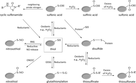
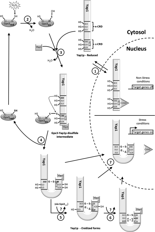
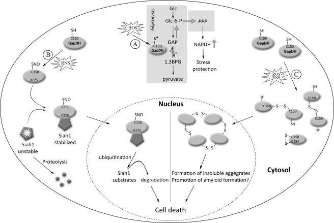
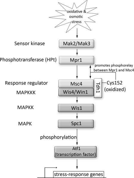
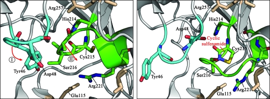
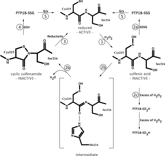
For many years, oxidative thiol modifications in cytosolic proteins were largely disregarded as in vitro artifacts, and considered unlikely to play significant roles within the reducing environment of the cell. Recent developments in in vivo thiol trapping technology combined with mass spectrometric analysis have now provided convincing evidence that thiol-based redox switches are used as molecular tools in many proteins to regulate their activity in response to reactive oxygen and nitrogen species. Reversible oxidative thiol modifications have been found to modulate the function of proteins involved in many different pathways, starting from gene transcription, translation and protein folding, to metabolism, signal transduction, and ultimately apoptosis. This review will focus on three well-characterized eukaryotic proteins that use thiol-based redox switches to influence gene transcription, metabolism, and signal transduction. The transcription factor Yap1p is a good illustration of how oxidative modifications affect the function of a protein without changing its activity. We use glyeraldehyde-3-phosphate dehydrogenase to demonstrate how thiol modification of an active site cysteine re-routes metabolic pathways and converts a metabolic enzyme into a pro-apoptotic factor. Finally, we introduce the redox-sensitive protein tyrosine phosphatase PTP1B to illustrate that reversibility is one of the fundamental aspects of redox-regulation.


Figures

FIG. 1.
FIG. 1.
Oxidative thiol modifications. Oxidation of cysteine thiol groups by H2O2 leads to sulfenic acid (R-SOH) formation. Sulfenic acids are either stabilized by nearby charges or react with neighboring thiols or proximal nitrogen to form disulfide bonds (R′-S-S-R″) or sulfenamide bonds (R′-S-NH-R′), respectively. In the presence of high H2O2 concentrations, overoxidation to sulfinic (R-SO2H) or sulfonic acid (R-SO3H) occurs. Although a few protein-specific sulfinic acid reductases have been identified, overoxidation is still considered to be largely irreversible in vivo. Alternatively, reaction of thiolate anions (RS-) with oxidized cysteines of other proteins or low molecular weight thiols such as glutathione (GSSG) leads to mixed disulfide bond formation (R′-S-S-R″) or S-glutathionylation (R-S-SG), respectively. Overoxidation of disulfide bonds in the presence of strong oxidants can cause thiosulfinate (R′-SO-S-R″) or irreversible thiosulfonate (R′-SO2-S-R″) formation. Most oxidative thiol modifications are reduced by members of the glutaredoxin (Grx) system and thioredoxin (Trx) system (reductants), which draw their reducing power from cellular NADPH. Exposure of thiolate anions to reactive nitrogen oxide species causes S-nitrosothiol formation, whereas treatment with peroxynitrite yields S-nitrothiol formation. The exact mechanism by which individual RNS cause oxidative thiol modifications in vivo is still under investigation.
FIG. 2.
FIG. 2.
Model of the Yap1p-Gpx3 redox relay. Under nonstress conditions, the cysteines of Yap1, which are clustered in two cysteine-rich domains (n-CRD, c-CRD), are reduced. (1) The nuclear export sequence (NES) of Yap1p is accessible for interaction with Crm1, and Yap1p shuttles between the cytosol and the nucleus. It prevents Yap1p's nuclear accumulation and guarantees low constitutive activity under nonstress conditions. (2) Upon exposure of yeast cells to oxidative stress conditions (i.e., H2O2) the active site cysteine (Cys36) of glutathione peroxidase Gpx3 attacks the O–O bond of H2O2, thereby forming a sulfenic acid at the active site cysteine. At the same time, Ybp1 binds to Yap1p. (3) In the next step, the active site sulfenic acid of Gpx reacts with Cys598 of Yap1p, causing the formation of a Gpx3-Yap1p disulfide intermediate. (4) A thiolate anion formed at Cys303 of Yap1p attacks the intermolecular disulfide bond, thereby causing the formation of the inter-domain Cys303–Cys598 disulfide bond and the recovery of reduced Gpx3. (5) Additional inter-domain disulfide bonds form in a process likely also involving Gpx3. While formation of the Cys310–Cys629 appears to increase transcriptional activation, (6) formation of Cys315–Cys620 might prolong the H2O2-mediated signal. The exact role of Ybp1 has yet to be determined, but it appears that Ybp1 mediates the redox signal from Gpx3 to Yap1p. (7) Disulfide bond formation appears to lead to conformational changes in Yap1p, which mask the NES and prevent nuclear export. Yap1p accumulates in the nucleus and activates the oxidative stress response. For reasons of simplicity, only one oxidized form of Yap1p is shown in the nucleus.
FIG. 3.
FIG. 3.
Oxidative thiol modifications of GapDH—From cytoprotection to cytotoxicity. (A) ROS-mediated inhibition of GapDH re-routes metabolic flux as immediate defense against oxidative insult. Oxidative stress-induced inhibition of GapDH as a central component of glycolysis leads to the rapid re-routing of glucose-6-phosphate into the pentose phosphate pathway (PPP), one of the key routes for NADPH production in the eukaryotic cytoplasm. NADPH serves as electron donor for glutathione reductase and thioredoxin reductase, and is therefore essential to restore and maintain the reducing environment of the cytosol. (B) Model of GapDH/Siah1-signaling cascade-dependent cell death. Exposure of GapDH to RNS causes S-nitrosation of Cys150 in GapDH. S-nitrosation stimulates the interaction between the E3-ubiquitin ligase Siah1 and GapDH. Siah1, which possesses a nuclear localization signal (NLS), escorts GapDH into the nucleus where the association between GapDH and Siah1 appears to stabilize the E3-ligase. This stabilization, in turn, increases the rate of degradation for a set of different target proteins, and ultimately triggers cell death. (C) Model of apoptosis triggered by ROS-mediated GapDH aggregation. Exposure of GapDH to excess ROS leads to the initial disulfide bond formation between either Cys150 and Cys154 (intramolecular disulfide bond) or between two active site cysteines Cys150. The latter leads to extensive conformational changes and exposes additional cysteines (e.g., Cys281) that undergo further intermolecular disulfide bond formation. This results in the accumulation of insoluble aggregates composed of multiple GapDH subunits in the cytosol and the nucleus. The aggregates are either intrinsically cytotoxic or interact with other aggregation-prone proteins (e.g., Aß, tau, α-synuclein) to cause cytotoxicity. For reasons of simplicity, only one subunit of the GapDH tetramer is shown.
FIG. 4.
FIG. 4.
Role of redox regulation by GapDH in stress signaling. In the fission yeast Schizosaccharomyces pombe, a multistep phosphorelay composed of the sensor kinase Mak2/Mak3, the histidine-containing phosphotransferase (HPt) Mpr1 and the Msc4 response regulator transmit stress signals, such as H2O2, to the Spc1 MAPK cascade. This cascade is composed of the Wis4/Win1 MAPKKKs, the MAPKK Wis1, and Spc1 MAPK as final receptor. Wis4/Win1 and Msc4 form a complex with the GapDH isoform Tdh1. H2O2-mediated oxidative modification of Tdh1's active site cysteine Cys152 enhances the interaction between Tdh1 and the Msc4 response regulator, which, in turn, promotes the interaction and phosphorelay signaling between the response regulator Msc4 and the HPt protein Mpr1. This interaction is required for the transmission of the H2O2 stress signal to Spc1.
FIG. 5.
FIG. 5.
Redox-mediated conformational changes in the catalytic site of PTP1B. Oxidation and sulfenamide bond formation of PTP1B's active site cysteine causes significant conformational changes in the catalytic site of PTP1B. Large rearrangements of both pTyr loop (blue) and PTP loop (green) inhibit substrate binding and apparently protect the active site Cys215 against irreversible overoxidation. Figures were made with PyMOL using the coordinates of reduced human PTP1B protein (2BGE) and the sulfenamide species (1OEM) deposited in the Protein Data Bank. (For interpretation of the references to color in this figure legend, the reader is referred to the web version of this article at www.liebertonline.com/ars).
FIG. 6.
FIG. 6.
PTP1B—Redox regulation by cyclic sulfenamide formation. Under nonstress conditions, the active site cysteine (Cys215) of tyrosine phosphatase PTP1B is reduced and PTP1B is active. (1) Upon exposure to oxidative stress conditions (e.g., H2O2), Cys215 becomes oxidized to sulfenic acid, which inactivates the enzyme. (2a) Reaction of the active site sulfenic acid with oxidized glutathione leads to S-glutathionylation (PTP1B-SSG). (2b) Alternatively, a nucleophilic attack of the backbone nitrogen of Ser216 on the Sγ-atom of Cys215 occurs, which results in the formation of a cyclic sulfenamide and the release of water. Sulfenamide formation is promoted by the environment of the catalytic site, with His214 playing a prominent role in polarizing the amide bond of Ser216. (2c) In the presence of excess H2O2, PTP1B's sulfenic acid is overoxidized to either sulfinic or sulfonic acid. (3) Active PTP1B can be either directly regenerated by reducing agents such as DTT in vitro or (4) indirectly via the reaction of cyclic sulfenamide with GSH and the formation of PTP1B-SSG. (5) Reduced active PTP1B is then formed by the subsequent reaction of PTP1B-SSG with the glutaredoxin (Grx) system.

References

    1. Abate C. Patel L. Rauscher FJ., 3rd Curran T. Redox regulation of fos and jun DNA-binding activity in vitro. Science. 1990;249:1157–1161. - PubMed
    1. Ahn SG. Thiele DJ. Redox regulation of mammalian heat shock factor 1 is essential for Hsp gene activation and protection from stress. Genes Dev. 2003;17:516–528. - PMC - PubMed
    1. Allen RG. Tresini M. Oxidative stress and gene regulation. Free Radic Biol Med. 2000;28:463–499. - PubMed
    1. Alonso A. Sasin J. Bottini N. Friedberg I. Friedberg I. Osterman A. Godzik A. Hunter T. Dixon J. Mustelin T. Protein tyrosine phosphatases in the human genome. Cell. 2004;117:699–711. - PubMed
    1. Aoyama K. Mitsubayashi Y. Aiba H. Mizuno T. Spy1, a histidine-containing phosphotransfer signaling protein, regulates the fission yeast cell cycle through the Mcs4 response regulator. J Bacteriol. 2000;182:4868–4874. - PMC - PubMed