. 2009 Jan 15;199(2):263-71.
doi: 10.1086/595735.

Perfusion abnormalities in children with cerebral malaria and malarial retinopathy

Affiliations

Perfusion abnormalities in children with cerebral malaria and malarial retinopathy

Nicholas A V Beare et al. J Infect Dis. .

Abstract

Background: In patients with cerebral malaria (CM), retinal angiography allows the study of infected central nervous system microvasculature in vivo. We aimed to examine retinal perfusion in children with CM by use of fluorescein angiography to investigate the pathophysiology of CM.

Methods: We performed fluorescein angiography on children with CM admitted to Queen Elizabeth Central Hospital, Malawi. We related angiograms to funduscopic findings.

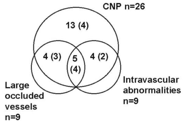
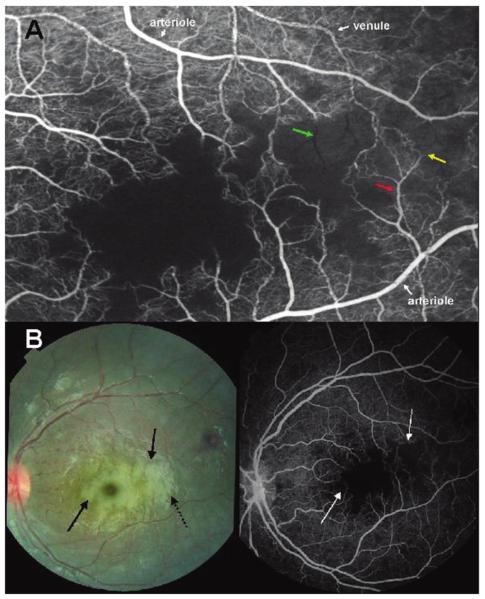
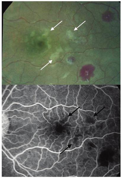
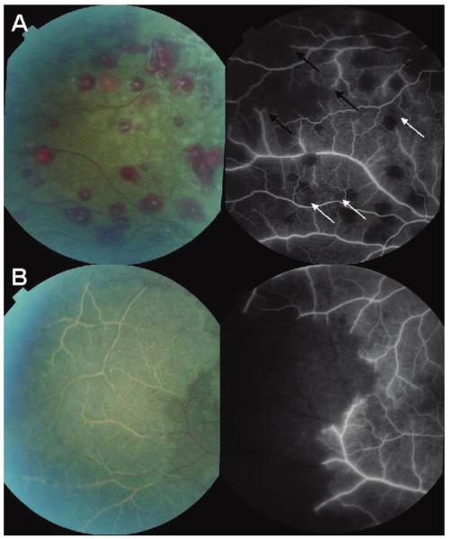
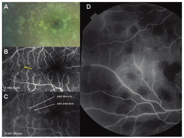
Results: Fluorescein angiography was performed for 34 patients with CM, and impaired perfusion was identified in 28 (82%). Areas of capillary nonperfusion (CNP) were seen in 26 patients (76%). Multiple, scattered areas of CNP were typical and topographically matched to retinal whitening. Larger retinal vessels were occluded in 9 patients (26%) who had associated ischemia. These vessels appeared white on ophthalmoscopy. Intravascular abnormalities were seen in 9 patients (26%), including filling defects and mottling of the blood column. Limited fluorescein leakage occurred in 15 patients (44%) and was not related to angiographic intravascular abnormalities or visible vessel discoloration.

Conclusions: Impaired perfusion occurs in the retinal microvasculature of most children with CM. This is evidence for hypoxia and ischemia as important components in the pathogenesis of CM. Vessel occlusion and filling defects are likely to be due to sequestration of infected erythrocytes. Interventions which improve perfusion or limit hypoxic injury may be beneficial in CM.


Figures

Figure 1
Figure 1
Venn diagram summarizing results of fluorescein angiograms performed for 34 patients. There were 28 patients with perfusion abnormalities. Large occluded vessels and intravascular abnormalities only occurred in the presence of capillary nonperfusion (CNP). There were 6 patients with no vascular abnormalities, and 2 patients with fluorescein leakage alone. The number in parentheses is the number with fluorescein leakage in each group.
Figure 2
Figure 2
A, Fluorescein angiogram image of the macula of a child with cerebral malaria showing large areas of capillary nonperfusion (center of picture). Arteriolar and venous circulation have been labelled to illustrate mottling of the blood column, which is most dense in the venules, but also occurs in the arterioles (red arrow). Vessel occlusion is widespread on the border of areas of nonperfusion (resulting in “pruning,” yellow arrow). “Ghost vessels'”can be seen (green arrow); these are nonperfused vessels, the contents of which are blocking the background choroidal fluorescence. B, Paired color retinal photograph and fluorescein angiogram of the same macula. The paired images show the topographical matching between areas of retinal whitening (solid black arrows) and areas of capillary nonperfusion (white arrows). The dark disc at the center of the largest area of macular whitening is the unaffected foveola. Dashed black arrow, artefactual glare overlying retinal whitening.
Figure 3
Figure 3
Paired color photograph and angiogram of the macula of a child with cerebral malaria. The images show the topographical matching between areas of retinal whitening (white arrows) and areas of capillary nonperfusion (black arrows). A retinal hemorrhage (bottom right, both images) demonstrates fluorescein masking or blocked fluorescence in the angiogram.
Figure 4
Figure 4
A, Paired color photograph and angiogram of the retinal periphery of a child with cerebral malaria. The angiogram image shows occlusion of retinal vessels (“pruning,”black arrows) with large associated areas of nonperfusion. Smaller areas of capillary nonperfusion are also illustrated (white arrows). B, Paired color photograph and angiogram of the retinal periphery of a different child. The child has extensive white retinal vessels within an area of confluent retinal whitening. On the angiogram, these white vessels are occluded and the whole area of retinal whitening is nonperfused. Both venules and arterioles are affected and are a normal red color up to the point of occlusion.
Figure 7
Figure 7
Color photograph (A) and early (B) and late (C) angiogram images of a macula showing leakage of fluorescein from terminal venules and not arterioles. This appears as fuzzy hyperfluorescence in C. The color image shows macular whitening around, and temporal to, the foveola (dark disc in the right of the image). There are corresponding areas of capillary nonperfusion (yellow arrow), which are associated with fluorescein leakage, indicating breakdown of the blood-retina barrier. D, Fluorescein image of peripheral retina showing leakage of fluorescein from veins in center of image. Round areas of hypofluorescence are all the result of fluorescence masking by retinal hemorrhages.

Similar articles

Cited by

References

    1. Bryce J, Boschi-Pinto C, Shibuya K, Black RE. WHO estimates of the causes of death in children. Lancet. 2005;365:1147–52. - PubMed
    1. Marsh K, Forster D, Waruiru C, et al. Indicators of life-threatening malaria in African children. N Engl J Med. 1995;332:1399–404. - PubMed
    1. Silamut K, Phu NH, Whitty C, et al. A quantitative analysis of the microvascular sequestration of malaria parasites in the human brain. Am J Pathol. 1999;155:395–410. - PMC - PubMed
    1. Lewallen S, White VA, Whitten RO, et al. Clinical-histopathological correlation of the abnormal retinal vessels in cerebral malaria. Arch Ophthalmol. 2000;118:924–8. - PubMed
    1. Seydel KB, Milner DA, Jr, Kamiza SB, Molyneux ME, Taylor TE. The distribution and intensity of parasite sequestration in comatose Malawian children. J Infect Dis. 2006;194:208–5. - PMC - PubMed

Publication types

MeSH terms