Review
. 2009 Jun;11(6):1289-99.
doi: 10.1089/ars.2008.2333.

Compartmentalization of redox signaling through NADPH oxidase-derived ROS

Affiliations
Review

Compartmentalization of redox signaling through NADPH oxidase-derived ROS

Masuko Ushio-Fukai. Antioxid Redox Signal. 2009 Jun.

Abstract

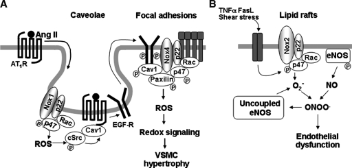
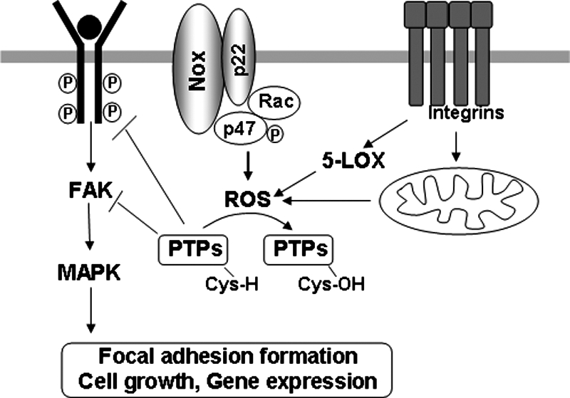
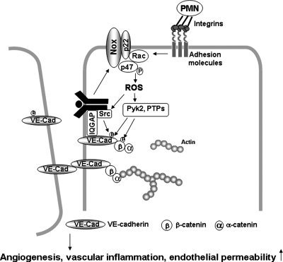
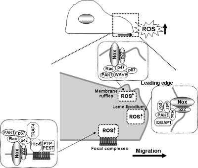
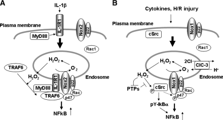
Reactive oxygen species (ROS) are generated in response to growth factors, cytokines, G protein-coupled receptor agonists, or shear stress, and function as signaling molecules in nonphagocytes. However, it is poorly understood how freely diffusible ROS can activate specific signaling, so-called "redox signaling." NADPH oxidases are a major source of ROS and now recognized to have specific subcellular localizations, and this targeting to specific compartments is required for localized ROS production. One important mechanism may involve the interaction of oxidase subunits with various targeting proteins localized in lamellipodial leading edge and focal adhesions/complexes. ROS are believed to inactivate protein tyrosine phosphatases, thereby establishing a positive-feedback system that promotes activation of specific redox signaling pathways involved in various functions. Additionally, ROS production may be localized through interactions of NADPH oxidase with signaling platforms associated with caveolae/lipid rafts, endosomes, and nucleus. These indicate that the specificity of ROS-mediated signal transduction may be modulated by the localization of Nox isoforms and their regulatory subunits within specific subcellular compartments. This review summarizes the recent progress on compartmentalization of redox signaling via activation of NADPH oxidase, which is implicated in cell biology and pathophysiologies.


Figures

FIG. 1.
FIG. 1.
Redox signaling in caveolae and lipid rafts. (A) In VSMCs, Ang II stimulation promotes AT1-receptor trafficking into caveolin-1–enriched membrane fractions where Nox1 is found. This causes localized ROS production and ROS-cSrc–dependent transactivation of the EGFR and its egress from caveolae. Tyrosine-phosphorylated EGFR and caveolin-1 subsequently appear at focal adhesions where Nox4 and paxillin localize, thereby forming redox signaling platforms. These events are essential for activation of specific redox signaling pathways involved in VSMC hypertrophy. (B) In ECs, stimulation with TNF-α, Fas ligand, and shear stress induces recruitments of Nox2, p47phox, and Rac1 into lipid rafts, thereby promoting raft-localized NADPH oxidase activation and ROS production and eNOS-derived NO within raft domains. This formation of redox signaling platforms in lipid rafts contributes to decrease in NO bioavailability and production of peroxynitrite, which uncouples NOS to produce more O2•-, which contributes to endothelial dysfunction.
FIG. 2.
FIG. 2.
Redox signaling at cell–matrix adhesions (focal adhesions). Growth factor activates NADPH oxidase, whereas integrin activation stimulates mitochondria and 5-lipoxygenase to generate localized ROS production at cell–matrix adhesions. ROS produced by synergistic action of integrins and growth factors induce oxidative inactivation of protein tyrosine phosphatases (PTPs), which negatively regulate RTK and FAK, thereby promoting downstream redox signaling events such as MAPK, leading to cell proliferation, survival, and gene expression.
FIG. 3.
FIG. 3.
Redox signaling at cell–cell contacts. PMN binding to ECs, inflammatory stimuli, or growth factors simulate localized ROS production via activation of NADPH oxidase at or near the cell–cell contacts in confluent ECs. ROS are involved in activation of Src, Pyk2, FAK, and PKC, or in oxidative inactivation of PTPs, thereby promoting tyrosine phosphorylation of VE-cadherin and β-catenin, which in turn promotes disruption of cell–cell contacts, and thus increasing endothelial permeability, migration, and proliferation, which are involved in angiogenesis.
FIG. 4.
FIG. 4.
Redox signaling in lamellipodial leading edge and focal complexes. Localized ROS signal at lamellipodial focal complexes is mediated through formation of p47phox-TRAF4-Hic5 complexes and oxidative inactivation of PTP-PEST by ROS, which is required for activation of Rac1 and its effector PAK1, which phosphorylate p47phox through formation of TRAF4-Rac1-PAK1 complexes. These p47phox–containing complexes create a positive-feedback loop to facilitate localized Nox-dependent ROS production, which contributes to directional cell migration. Localized ROS at membrane ruffles and lamellipodial leading edges are mediated through p47phox-WAVE1-Rac1-PAK1 complexes, which phosphorylate p47phox as well as Rac1-IQGAP1-Nox2 targeting to the leading edge through the scaffolding function of IQGAP1, respectively. These NADPH oxidase–targeting mechanisms are required for ROS-dependent directional cell migration.
FIG. 5.
FIG. 5.
Redox signaling in endosomes. (A) Binding of IL-1 to IL-1R1 on the plasma membrane promotes MyD88 association with IL-1R1, which triggers endocytosis of the IL-1R1-MyD88 complex and subsequent recruitment of Rac1 and Nox2 into the endosomal compartment. Localized ROS signal in endosomes facilitates the redox-dependent association of TRAF6 with the receptor complex, which contributes to activation of NF-κB. (B) Hypoxia/reoxygenation (H/R) injury induces endosomal recruitment of cSrc and Rac1, thereby activating Nox1-dependent ROS production and its downstream cSrc, which phosphorylates Iκ-Bα, and thus leading to NF-κB activation. In VSMCs, Nox1 colocalizes with ClC-3 in endosomes, which is required for cytokine-induced ROS production within endosomes and its downstream NF-κB activation.

Similar articles

Cited by

References

    1. Abate C. Patel L. Rauscher FJ., 3rd Curran T. Redox regulation of fos and jun DNA-binding activity in vitro. Science (New York) 1990;249:1157–1161. - PubMed
    1. Abid MR. Kachra Z. Spokes KC. Aird WC. NADPH oxidase activity is required for endothelial cell proliferation and migration. FEBS Lett. 2000;486:252–256. - PubMed
    1. Allen RG. Tresini M. Oxidative stress and gene regulation. Free Radic Biol Med. 2000;28:463–499. - PubMed
    1. Allingham MJ. van Buul JD. Burridge K. ICAM-1-mediated, Src- and Pyk2-dependent vascular endothelial cadherin tyrosine phosphorylation is required for leukocyte transendothelial migration. J Immunol. 2007;179:4053–4064. - PubMed
    1. Ambasta RK. Kumar P. Griendling KK. Schmidt HH. Busse R. Brandes RP. Direct interaction of the novel Nox proteins with p22phox is required for the formation of a functionally active NADPH oxidase. J Biol Chem. 2004;279:45935–45941. - PubMed

Publication types

LinkOut - more resources