

Delivery of rapamycin to dendritic cells using degradable microparticles
- PMID: 19000726
- PMCID: PMC2925512
- DOI: 10.1016/j.jconrel.2008.10.011
Delivery of rapamycin to dendritic cells using degradable microparticles
Abstract
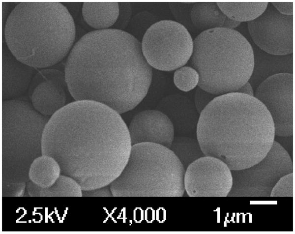
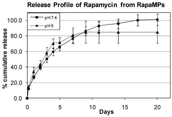
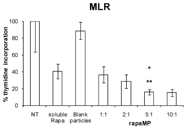
Degradable microparticles have the potential to protect and release drugs over extended periods and, if sized appropriately, can be passively targeted to phagocytic cells in vivo. Dendritic cells (DC) are a class of phagocytic cells known to play important roles in transplant rejection. Previously, we have demonstrated that DC treated with an immunosuppressive drug, rapamycin, have the ability to suppress transplant rejection. Herein, we describe a strategy to deliver an intracellular depot of rapamycin to DC. To achieve this, rapamycin was encapsulated into ~3.4 microm sized poly(lactic-co-glycolic)acid (PLGA) microparticles (rapaMPs), and release behavior was examined under intra-phagosomal (pH=5) and extracellular (pH=7.4) conditions. It was observed that 4 days following phagocytosis of rapaMP, DC have significantly reduced ability to activate T cells, in comparison to DC treated with soluble rapamycin. Hence, we conclude that DC-specific intracellular delivery of rapamycin results in better efficacy of the drug, with respect to its ability to modulate DC function, when compared to treating DC with extracellular rapamycin.
Figures
Similar articles
-
Delivery of rapamycin by PLGA nanoparticles enhances its suppressive activity on dendritic cells.J Biomed Mater Res A. 2008 Mar 15;84(4):885-98. doi: 10.1002/jbm.a.31373. J Biomed Mater Res A. 2008. PMID: 17647224
-
Rapamycin-conditioned dendritic cells are poor stimulators of allogeneic CD4+ T cells, but enrich for antigen-specific Foxp3+ T regulatory cells and promote organ transplant tolerance.J Immunol. 2007 Jun 1;178(11):7018-31. doi: 10.4049/jimmunol.178.11.7018. J Immunol. 2007. PMID: 17513751
-
Spray-dried chitosan microparticles for cellular delivery of an antigenic protein: physico-chemical properties and cellular uptake by dendritic cells and macrophages.Pharm Res. 2013 Jun;30(6):1677-97. doi: 10.1007/s11095-013-1014-7. Epub 2013 Mar 13. Pharm Res. 2013. PMID: 23483441
-
Activation of antigen-specific T cell-responses by mannan-decorated PLGA nanoparticles.Pharm Res. 2011 Sep;28(9):2288-301. doi: 10.1007/s11095-011-0459-9. Epub 2011 May 11. Pharm Res. 2011. PMID: 21560020
-
Rational design of nanoparticles towards targeting antigen-presenting cells and improved T cell priming.J Control Release. 2017 Jul 28;258:182-195. doi: 10.1016/j.jconrel.2017.05.014. Epub 2017 May 13. J Control Release. 2017. PMID: 28511928
Cited by
-
Polymeric micro- and nanoparticles for immune modulation.Biomater Sci. 2018 Dec 18;7(1):14-30. doi: 10.1039/c8bm01285g. Biomater Sci. 2018. PMID: 30418444 Free PMC article. Review.
-
Nano and Microparticle Emerging Strategies for Treatment of Autoimmune Diseases: Multiple Sclerosis and Type 1 Diabetes.Adv Healthc Mater. 2020 Jun;9(11):e2000164. doi: 10.1002/adhm.202000164. Epub 2020 Apr 27. Adv Healthc Mater. 2020. PMID: 32519501 Free PMC article. Review.
-
Solid Lipid Nanoparticles (SLNs) for Intracellular Targeting Applications.J Vis Exp. 2015 Nov 17;(105):53102. doi: 10.3791/53102. J Vis Exp. 2015. PMID: 26650036 Free PMC article.
-
Biomedical Applications of Biodegradable Polymers.J Polym Sci B Polym Phys. 2011 Jun 15;49(12):832-864. doi: 10.1002/polb.22259. J Polym Sci B Polym Phys. 2011. PMID: 21769165 Free PMC article.
-
Micro and nanoparticle drug delivery systems for preventing allotransplant rejection.Clin Immunol. 2015 Sep;160(1):24-35. doi: 10.1016/j.clim.2015.04.013. Epub 2015 May 1. Clin Immunol. 2015. PMID: 25937032 Free PMC article. Review.
References
-
- Tantravahi J, Womer KL, Kaplan B. Why hasn't eliminating acute rejection improved graft survival? Annual Review of Medicine. 2007;58:369–385. - PubMed
-
- Lechler RI, Sykes M, Thomson AW, Turka LA. Organ Transplantation - How much of the promise has been realized. Nature Medicine. 2005;11(6):605–613. - PubMed
-
- Banchereau J, Steinman RM. Dendritic cells and the control of immunity. Nature. 1998;392:245–252. - PubMed
-
- Steinman RM, Hawiger D, Nussenzweig MC. Tolerogenic Dendritic Cells. Annual Review of Immunology. 2003;21:685–711. - PubMed
-
- Krämer BK, Böger C, Krüger B, Marienhagen J, Pietrzyk M, Obed A, Paczek L, Mack M, Banas B. Cardiovascular Risk Estimates and Risk Factors in Renal Transplant Recipients. Transplantation Proceedings. 2005;37:1868–1870. - PubMed
Publication types
MeSH terms
Substances
Grants and funding
LinkOut - more resources
Full Text Sources
Other Literature Sources
