Molecular mechanisms regulating glucocorticoid sensitivity and resistance
- PMID: 19000736
- PMCID: PMC2674248
- DOI: 10.1016/j.mce.2008.10.001
Molecular mechanisms regulating glucocorticoid sensitivity and resistance
Abstract
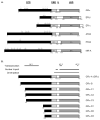
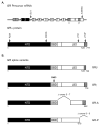
Glucocorticoid receptor agonists are mainstays in the treatment of various malignancies of hematological origin. Glucocorticoids are included in therapeutic regimens for their ability to stimulate intracellular signal transduction cascades that culminate in alterations in the rate of transcription of genes involved in cell cycle progression and programmed cell death. Unfortunately, subpopulations of patients undergoing systemic glucocorticoid therapy for these diseases are or become insensitive to glucocorticoid-induced cell death, a phenomenon recognized as glucocorticoid resistance. Multiple factors contributing to glucocorticoid resistance have been identified. Here we summarize several of these mechanisms and describe the processes involved in generating a host of glucocorticoid receptor isoforms from one gene. The potential role of glucocorticoid receptor isoforms in determining cellular responsiveness to glucocorticoids is emphasized.
Figures


Similar articles
-
Tissue-specific glucocorticoid action: a family affair.Trends Endocrinol Metab. 2008 Nov;19(9):331-9. doi: 10.1016/j.tem.2008.07.009. Epub 2008 Sep 19. Trends Endocrinol Metab. 2008. PMID: 18805703 Free PMC article. Review.
-
Glucocorticoid Resistance: Interference between the Glucocorticoid Receptor and the MAPK Signalling Pathways.Int J Mol Sci. 2021 Sep 17;22(18):10049. doi: 10.3390/ijms221810049. Int J Mol Sci. 2021. PMID: 34576214 Free PMC article. Review.
-
Mechanisms of Brain Glucocorticoid Resistance in Stress-Induced Psychopathologies.Biochemistry (Mosc). 2017 Mar;82(3):351-365. doi: 10.1134/S0006297917030142. Biochemistry (Mosc). 2017. PMID: 28320277 Review.
-
Corticosteroids: Mechanisms of Action in Health and Disease.Rheum Dis Clin North Am. 2016 Feb;42(1):15-31, vii. doi: 10.1016/j.rdc.2015.08.002. Rheum Dis Clin North Am. 2016. PMID: 26611548 Free PMC article. Review.
-
Exploring the molecular mechanisms of glucocorticoid receptor action from sensitivity to resistance.Endocr Dev. 2013;24:41-56. doi: 10.1159/000342502. Epub 2013 Feb 1. Endocr Dev. 2013. PMID: 23392094 Free PMC article. Review.
Cited by
-
Ectopic microRNA-150-5p transcription sensitizes glucocorticoid therapy response in MM1S multiple myeloma cells but fails to overcome hormone therapy resistance in MM1R cells.PLoS One. 2014 Dec 4;9(12):e113842. doi: 10.1371/journal.pone.0113842. eCollection 2014. PLoS One. 2014. PMID: 25474406 Free PMC article.
-
Expression, regulation and function of phosphofructo-kinase/fructose-biphosphatases (PFKFBs) in glucocorticoid-induced apoptosis of acute lymphoblastic leukemia cells.BMC Cancer. 2010 Nov 23;10:638. doi: 10.1186/1471-2407-10-638. BMC Cancer. 2010. PMID: 21092265 Free PMC article.
-
Research resource: transcriptional response to glucocorticoids in childhood acute lymphoblastic leukemia.Mol Endocrinol. 2012 Jan;26(1):178-93. doi: 10.1210/me.2011-1213. Epub 2011 Nov 10. Mol Endocrinol. 2012. PMID: 22074950 Free PMC article.
-
A substitution in the ligand binding domain of the porcine glucocorticoid receptor affects activity of the adrenal gland.PLoS One. 2012;7(9):e45518. doi: 10.1371/journal.pone.0045518. Epub 2012 Sep 18. PLoS One. 2012. PMID: 23029068 Free PMC article.
-
Inhibition of histone deacetylase 2 expression by elevated glucocorticoid receptor beta in steroid-resistant asthma.Am J Respir Crit Care Med. 2010 Oct 1;182(7):877-83. doi: 10.1164/rccm.201001-0015OC. Epub 2010 Jun 10. Am J Respir Crit Care Med. 2010. PMID: 20538962 Free PMC article.
References
-
- Alarid ET. Lives and times of nuclear receptors. Mol Endocrinol. 2006;20:1972–1981. - PubMed
-
- Antakly T, Thompson EB, O’Donnell D. Demonstration of the intracellular localization and up-regulation of glucocorticoid receptor by in situ hybridization and immunocytochemistry. Cancer Res. 1989;49:2230s–2234s. - PubMed
-
- Ashraf J, Kunapuli S, Chilton D, Thompson EB. Cortivazol mediated induction of glucocorticoid receptor messenger ribonucleic acid in wild-type and dexamethasone-resistant human leukemic (CEM) cells. J Steroid Biochem Mol Biol. 1991;38:561–568. - PubMed
-
- Ashraf J, Thompson EB. Identification of the activation-labile gene: a single point mutation in the human glucocorticoid receptor presents as two distinct receptor phenotypes. Mol Endocrinol. 1993;7:631–642. - PubMed
Publication types
MeSH terms
Substances
Grants and funding
LinkOut - more resources
Full Text Sources
Medical

