. 2008 Nov 12;16(11):1649-58.
doi: 10.1016/j.str.2008.08.013.

Core structure of the yeast spt4-spt5 complex: a conserved module for regulation of transcription elongation

Affiliations

Core structure of the yeast spt4-spt5 complex: a conserved module for regulation of transcription elongation

Min Guo et al. Structure. .

Abstract

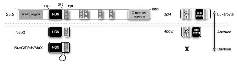
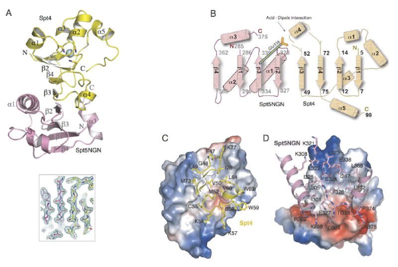
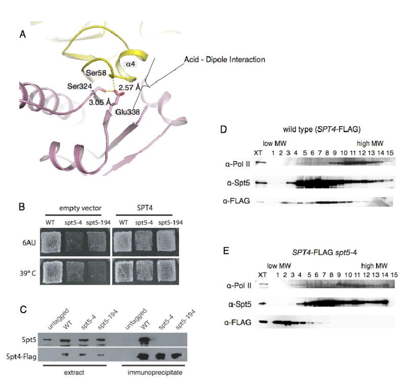
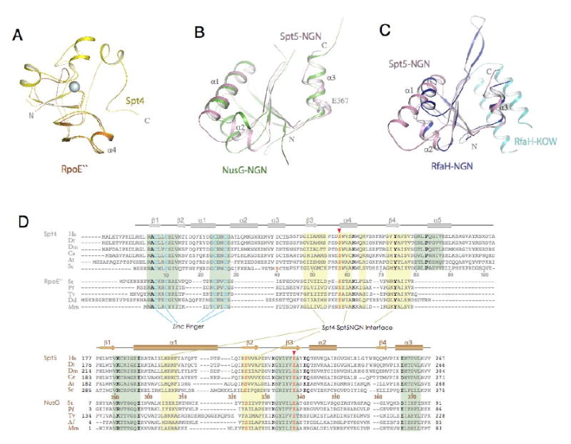
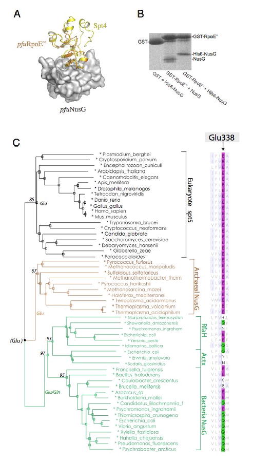
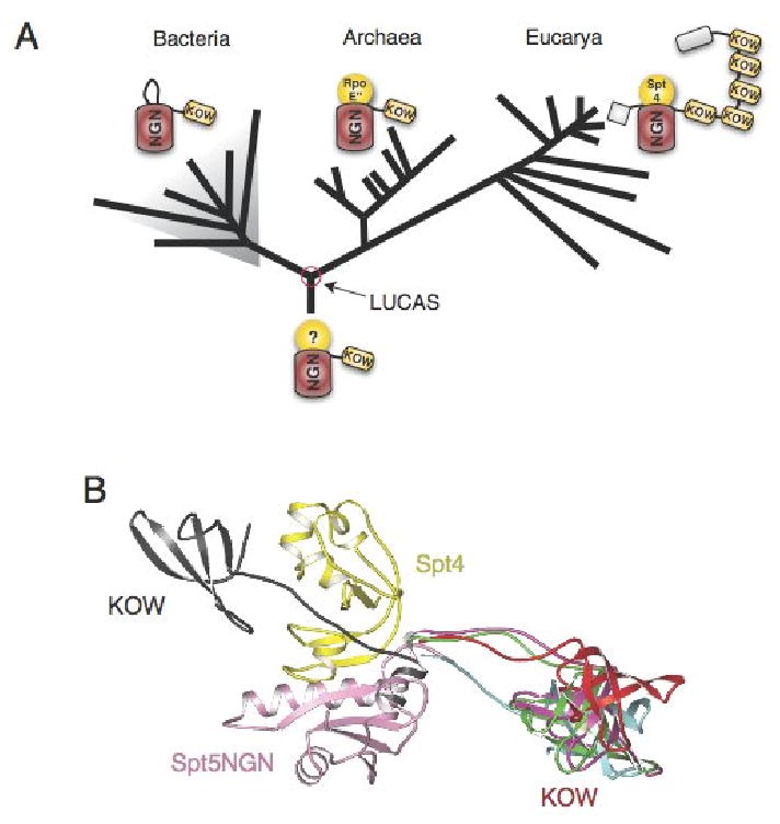
The Spt4-Spt5 complex is an essential RNA polymerase II elongation factor found in all eukaryotes and important for gene regulation. We report here the crystal structure of Saccharomyces cerevisiae Spt4 bound to the NGN domain of Spt5. This structure reveals that Spt4-Spt5 binding is governed by an acid-dipole interaction between Spt5 and Spt4. Mutations that disrupt this interaction disrupt the complex. Residues forming this pivotal interaction are conserved in the archaeal homologs of Spt4 and Spt5, which we show also form a complex. Even though bacteria lack a Spt4 homolog, the NGN domains of Spt5 and its bacterial homologs are structurally similar. Spt4 is located at a position that may help to maintain the functional conformation of the following KOW domains in Spt5. This structural and evolutionary perspective of the Spt4-Spt5 complex and its homologs suggest that it is an ancient, core component of the transcription elongation machinery.


Figures

Figure 1
Figure 1. Linear maps of the eukaryotic Spt4, Spt5 and the prokaryotic NusG, RpoE″
Conserved motifs are shown as boxes. The C-terminal portion of Spt4 is absent in RpoE″. Residue numbers refer to Saccharomyces cerevisiae Spt5.
Figure 2
Figure 2. Architecture of Spt4-Spt5NGN complex
(A) Ribbon diagram of the structure of Spt4-Spt5NGN complex. The Spt4 and Spt5 NGN domains are shown in yellow and pink, respectively; the Zn atom is shown as a blue sphere. The central β-sheet is shown by the 2mfo-Dfc map calculated with refmac coefficients contoured at 2σ level. (B) Topology of the secondary structure elements. Residue numbers and the N and C termini of Spt4 and Spt5NGN are indicated. (C, D) Views of the Spt4-Spt5NGN interface. C, Surface of Spt5NGN is shown and colored according to electrostatic, interacting residues in Spt4 are labeled and shown as yellow sticks; D, Surface of Spt4 and interacting residues in Spt5NGN are shown, interacting residues in Spt5NGN are labeled and shown as pink sticks.
Figure 3
Figure 3. Mutations of the acid-dipole interaction disrupt the Spt4-Spt5 complex
(A) Conserved acid--α-helix dipole interaction between Helix_α4 of Spt4 and Glu338 of Spt5NGN. The carboxyl of Glu338 is also fixed by two hydrogen-bonds with Spt4_Ser58 and Spt5_Ser324. The coloring is the same as in Figure 2. (B) Suppression of spt5-4 and spt5-194 by overexpression of Spt4. The indicated strains were transformed with either empty vector or a high copy Spt4 plasmid and replica plated to YPD media at 39° C or SC-ura media with 50 microgram per ml 6-azauracil at 30° C. (C) Co-immunoprecipitation of Spt4-Spt5 from extracts of Spt5, Spt5-4 or Spt5-194 cells. (D, E) The spt5-4 mutation disrupts the Spt4-Spt5 complex; Extracts of cells expressing wild type Spt5 or Spt5-4 and flag-tagged Spt4 were fractionated on 10-30% sucrose gradients. Fractions were separated on SDS gels and blotted with the indicated anti-sera.
Figure 4
Figure 4. Structural conservation of Spt4 and Spt5NGN domain
(A) Structural superimposition of Spt4 (yellow) with Pyrococcus furiosus RpoE″ (orange, pdb1ryq). (B) Structural superimposition of Spt5NGN domain with the Aquifex aeolius NusG (green, pdb1npp). (C) structural superimposition of Spt5NGN domain with the e.coli RfaH (pdb2oug, NGN in dark blue and KOW in light blue). (D) Alignment of representative Spt4 and Spt5 homologues. Residues that fall in the binding interface between Spt4 and the Spt5NGN domain are noted with a yellow background; residues that contribute to structural stability are noted by the green background. Highly conserved residues are depicted in bold type; residues in pink are involved in the acid-dipole interaction. Abbreviations: Hs, Homo sapiens; Dr, Danio rerio; Dm, Drosophila melanogaster; Ce, Caenorhabditis elegans; At, Arabidopsis thaliana; Sc, Saccharomyces cerevisiae; St, Sulfolobus tokodaii; Pf, Pyrococcus furiosus; Tv, Thermoplasma volcanium; Dd, Dictyostelium discoideum; Af, Archaeoglobus fulgidus; Mm, Methanococcus maripaludis;
Figure 5
Figure 5. Conservation of the Spt4-Spt5 Interface
(A) Model of Archaeal NusG bound to RpoE″ : Archaeal RpoE″-NusG complex was modeled by superimposing the pfuRpoE″ structure and pfuNusG model onto the Spt4-Spt5NGN structure. The pfuNusG model is shown as a white surface; yellow, Spt4; orange, pfuRpoE″. (B) GST Pull-down assay of Methanocaldococcus jannaschii RpoE″-NusG interaction. (C) A sequence-based phylogeny derived from an alignment of NGN domain in Spt5/NusG homologues. Organism names are color-coded according to the domain of life. Alignment of Glu338 for the acid-dipole interaction is shown at right. Bootstrap support is indicated for major braches.
Figure 6
Figure 6. Evolution of the Spt4-Spt5 Complex
(A) An evolutionary model of Spt4-Spt5 shows the distribution and domain organization of their homologues across evolution. (B) Superimposition of 5 distinct bacterial aaeNusG structures(described in Reay et al., 2004) and the eukaryotic Spt4-Spt5NGN complex. Only the Spt4-Spt5NGN and KOW structures are shown, the superimposed NusG_NGN domains were omitted for clarity. A notable feature of the NusG structure is the flexible linkage between NGN and KOW domains. In one of the 5 NusG structures shown here (in black, pdb1npp, chain D), the orientation of the KOW domain relative to the NGN domain is dramatically different from that of the other 4 structures.

References

    1. Allison LA, Moyle M, Shales M, Ingles CJ. Extensive homology among the largest subunits of eukaryotic and prokaryotic RNA polymerases. Cell. 1985;42:599–610. - PubMed
    1. Andrulis ED, Guzman E, Doring P, Werner J, Lis JT. High-resolution localization of Drosophila Spt5 and Spt6 at heat shock genes in vivo: roles in promoter proximal pausing and transcription elongation. Genes & development. 2000;14:2635–2649. - PMC - PubMed
    1. Artsimovitch I, Landick R. The transcriptional regulator RfaH stimulates RNA chain synthesis after recruitment to elongation complexes by the exposed nontemplate DNA strand. Cell. 2002;109:193–203. - PubMed
    1. Barton NH, Briggs DEG, Eisen JA, Goldstein DB, Patel NH. Evolution. Cold Spring Harbor Laboratory Press; 2007. Evolution; pp. 109–128.
    1. Basrai MA, Kingsbury J, Koshland D, Spencer F, Hieter P. Faithful chromosome transmission requires Spt4p a putative regulator of chromatin structure in Saccharomyces cerevisiae. Mol Cell Biol. 1996;16:2838–2847. - PMC - PubMed

Publication types

MeSH terms

Substances

Associated data