

Inherited mitochondrial optic neuropathies
- PMID: 19001017
- PMCID: PMC2643051
- DOI: 10.1136/jmg.2007.054270
Inherited mitochondrial optic neuropathies
Erratum in
- J Med Genet. 2011 Apr;48(4):284
Abstract
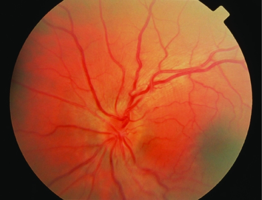
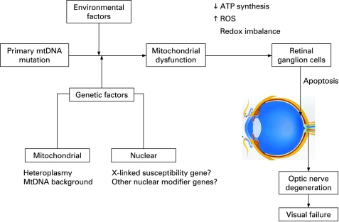
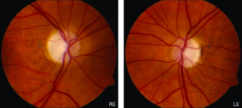
Leber hereditary optic neuropathy (LHON) and autosomal dominant optic atrophy (DOA) are the two most common inherited optic neuropathies and they result in significant visual morbidity among young adults. Both disorders are the result of mitochondrial dysfunction: LHON from primary mitochondrial DNA (mtDNA) mutations affecting the respiratory chain complexes; and the majority of DOA families have mutations in the OPA1 gene, which codes for an inner mitochondrial membrane protein critical for mtDNA maintenance and oxidative phosphorylation. Additional genetic and environmental factors modulate the penetrance of LHON, and the same is likely to be the case for DOA which has a markedly variable clinical phenotype. The selective vulnerability of retinal ganglion cells (RGCs) is a key pathological feature and understanding the fundamental mechanisms that underlie RGC loss in these disorders is a prerequisite for the development of effective therapeutic strategies which are currently limited.
Conflict of interest statement
Figures
References
-
- Schaefer AM, McFarland R, Blakely EL, He L, Whittaker RG, Taylor RW, Chinnery PF, Turnbull DM. Prevalence of mitochondrial DNA disease in adults. Ann Neurol 2008;63:35–9 - PubMed
-
- Leber T. Ueber hereditaere und congenital angelegte sehnervenleiden. Graefes Arch Opthal 1871;17:249–91
-
- Bell J. Hereditary optic atrophy (Leber’s disease). Pearson K, ed. The treasury of human inheritance Cambridge: Cambridge University Press, 1931:345–423
-
- Imai Y, Moriwaki D. A probable case of cytoplasmic inheritance in man: a critique of Leber’s disease. J Genet 1936;33:163–7
Publication types
MeSH terms
Substances
Grants and funding
LinkOut - more resources
Full Text Sources
Other Literature Sources
Medical
Molecular Biology Databases