. 2008 Nov 25;118(22):2225-34.
doi: 10.1161/CIRCULATIONAHA.108.788067. Epub 2008 Nov 10.

Proarrhythmic defects in Timothy syndrome require calmodulin kinase II

Affiliations

Proarrhythmic defects in Timothy syndrome require calmodulin kinase II

William H Thiel et al. Circulation. .

Abstract

Background: Timothy syndrome (TS) is a disease of excessive cellular Ca(2+) entry and life-threatening arrhythmias caused by a mutation in the primary cardiac L-type Ca(2+) channel (Ca(V)1.2). The TS mutation causes loss of normal voltage-dependent inactivation of Ca(V)1.2 current (I(Ca)). During cellular Ca(2+) overload, the calmodulin-dependent protein kinase II (CaMKII) causes arrhythmias. We hypothesized that CaMKII is a part of the proarrhythmic mechanism in TS.

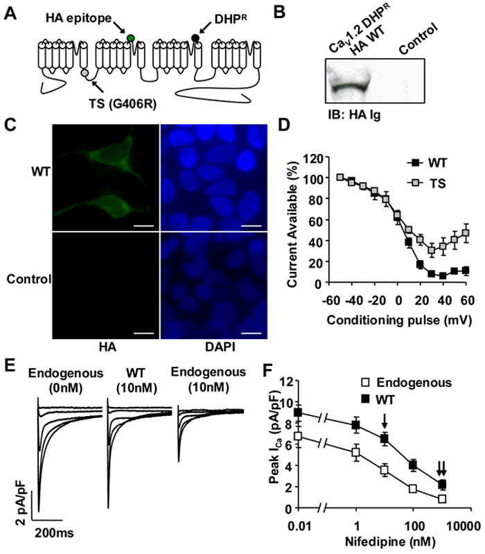
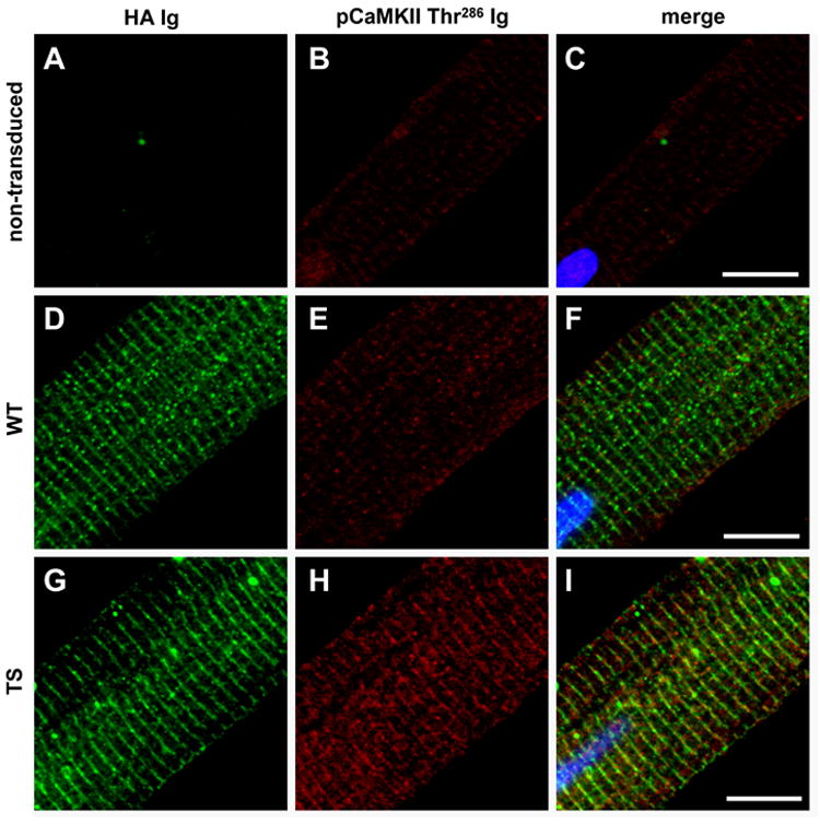
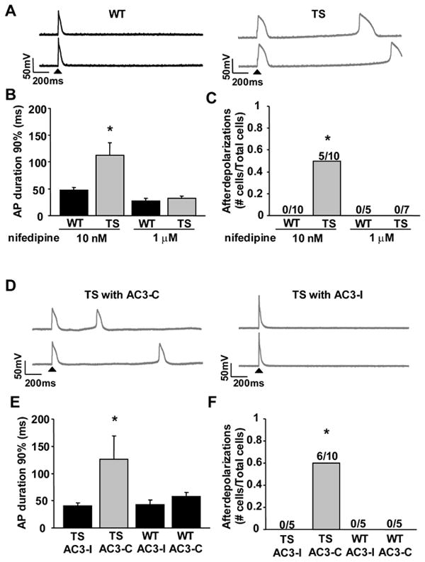
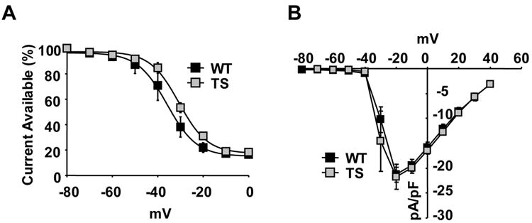
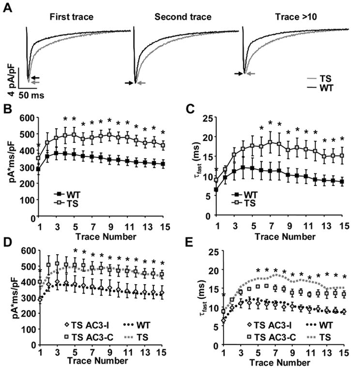
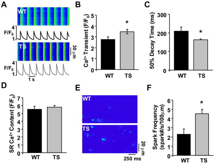
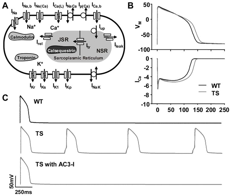
Methods and results: We developed an adult rat ventricular myocyte model of TS (G406R) by lentivirus-mediated transfer of wild-type and TS Ca(V)1.2. The exogenous Ca(V)1.2 contained a mutation (T1066Y) conferring dihydropyridine resistance, so we could silence endogenous Ca(V)1.2 with nifedipine and maintain peak I(Ca) at control levels in infected cells. TS Ca(V)1.2-infected ventricular myocytes exhibited the signature voltage-dependent inactivation loss under Ca(2+) buffering conditions, not permissive for CaMKII activation. In physiological Ca(2+) solutions, TS Ca(V)1.2-expressing ventricular myocytes exhibited increased CaMKII activity and a proarrhythmic phenotype that included action potential prolongation, increased I(Ca) facilitation, and afterdepolarizations. Intracellular dialysis of a CaMKII inhibitory peptide, but not a control peptide, reversed increases in I(Ca) facilitation, normalized the action potential, and prevented afterdepolarizations. We developed a revised mathematical model that accounts for CaMKII-dependent and CaMKII-independent effects of the TS mutation.

Conclusions: In TS, the loss of voltage-dependent inactivation is an upstream initiating event for arrhythmia phenotypes that are ultimately dependent on CaMKII activation.


Conflict of interest statement

Conflict of Interest Disclosures none

Figures

Figure 1
Figure 1
Dihydropyridine-resistant CaV1.2 α subunit Timothy Syndrome (TS) model. (A) A topology diagram of CaV1.2 depicting dihydropyridine resistance mutation (DHPR, black circle), extracellular hemaglutanin epitope (HA, green circle) and the TS mutation (G406R, gray circle) on the I-II intracellular loop. (B) Immunoblot (HA Ig) of HEK293T cells expressing the modified CaV1.2 or empty vector control. (C) FITC immunofluorescence (HA Ig) of HEk293T cells expressing the modified CaV1.2 with corresponding nuclear stain by DAPI (Scale bar, 10μm). (D) CaV1.2 TS expressing HEK293T cells show a reduction in VDI as compared to HEK293T cells transfected with CaV1.2 WT (N=5 cells/point). (E) Exogenous CaV1.2 with dihydropyridine mutation is resistant to nifedipine. (F) Preserved ICa during exposure to nifedipine. The single arrow indicates the nifedipine concentration (10nM) used to study the cellular consequences of the TS mutation, and the double arrow indicates the nifedipine concentration (1μM) to overcome dihydropyridine resistance and block the majority of ICa (N=5-8 cells/point, P<0.05 at each nifedipine concentration).
Figure 2
Figure 2
CaMKII recruitment in the TS adult ventricular myocyte model. (A-C) Non-transduced, (D-F) WT and (G-I) TS adult ventricular myocytes (field stimulated 1Hz for 5 minutes in Tyrodes with 1.8mM CaCl2). (A,D,G) Exogenous CaV1.2 channels are expressed in regularly distributed punctae across ventricular myocytes as shown by HA immunostaining. Both WT and TS CaV1.2 show spacing consistent with T-tubule network localization. HA immunofluorescence section of CaV1.2 WT, TS mutation, and uninfected negative control. (H) More activated CaMKII (pCaMKII Thr286) immuno-stained with (I) TS ventricular myocytes as compared to WT (E and F) and non-transduced (B and C) ventricular myocytes. (Scale bar, 10μm)
Figure 3
Figure 3
CaMKII inhibition reverses TS ventricular myocyte action potential (AP) prolongation and afterdepolarizations. (A) Action potential recordings from WT and TS ventricular myocytes. The first action potential for each sweep was initiated by injected current (arrow head), but the subsequent action potentials in TS arose from spontaneous afterdepolarizations. (B) CaV1.2 TS results in an increased action potential duration (N=5-10 cells/group, *P=0.018) and (C) afterdepolarizations (N=5-10 cells/group, *P=0.033). Numerals indicate the fraction of cells studied with afterdepolarizations. 1μm Nifedipine inhibited a majority of ICa and so prevented the CaV1.2 TS increase in (B) action potential duration (N=5-7 cells/group, P-0.39) and (C) frequency of afterdepolarizations (N=5-7 cells/group, P=1.0). (D) Action potential recordings from TS ventricular myocytes with either the CaMKII inhibitory peptide, AC3-I, or a control peptide, AC3-C. (E and F) Dialyzing AC3-I restored action potential duration in TS to WT levels and prevented afterdepolarizations (N=5-10 cells/group, TS with AC3-I compared to WT: APD90% P=0.403, afterdepolarizations P=1.0). AC3-I resulted in a non-significant shortening of the WT AP duration (N=5 cells/group, P=0.25) and no significant change in afterdepolarizations (N=5 cells/group, P=1.0) compared to WT with no peptide. (D-F) Dialyzing the control peptide, AC3-C, did not alter the TS mutation affects on action potential duration or afterdepolarizations (N=5-10 cells/group, TS with AC3-C compared to TS with AC3-I: APD90% *P=0.017, afterdepolarizations *P=0.044). AC3-C did not alter the AP duration (N=5 cells/group, P=0.28) or afterdepolarizations (N=5 cells/group, P=1.0) of WT with no peptide.
Figure 4
Figure 4
TS mutation shifts the VDI independent of Ca2+ signaling. (A) The TS mutation shifts the CaV1.2 IBa VDI (N=5 cells/point, *P=0.008), (B) without changing the current-voltage (IV) relationship (N=5 cells/group, P=0.88).
Figure 5
Figure 5
TS mutation enhances ICa facilitation. (A) TS ventricular myocytes exhibit increased peak ICa (arrows) during the first depolarizing voltage clamp command step (-80mV to 0mV, 300ms, 0.5Hz) and slowing of inactivation during all depolarizing steps. (B) Integrated ICa evoked by repetitive depolarizing voltage command steps (as in A above) is greater in TS mutation than WT (N=6-7 cells/point, ANOVA P<0.001, * P<0.05). (C) The time constant of the fast component of ICa inactivation (τfast) is significantly slower in TS ventricular myocytes than WT (N=6-7 cells/point, ANOVA P<0.001, * P<0.05). (D and E) Integrated ICa and τfast were restored to WT levels in TS ventricular myocytes dialyzed with the CaMKII inhibitory peptide, AC3-I (N=5-6 cells/point, TS with AC3-I compared to WT: integrated ICa ANOVA P=0.522, τfast ANOVA P=0.294). Dialyzing the control peptide, AC3-C, did not alter the TS mutation affects on ICa facilitation (N=5 cells/group, TS with AC3-C compared to TS with AC3-I: integrated ICa ANOVA P<0.001, * P<0.05; τfast ANOVA P<0.001, * P<0.05).
Figure 6
Figure 6
The TS mutation augments intracellular Ca2+ handling. (A) Confocal Ca2+ transient recordings from WT and TS ventricular myocytes. (B) Summary data showing TS mutation causes an increase in the peak Ca2+ transient during 1Hz stimulations (N=14-28 cells/group, *P=0.042). (C) Summary data showing the 50% decay time of the whole cell Ca2+ transients were faster in TS ventricular myocytes (N=14-28 cells/group, *P=0.047). (D) No difference was observed in SR Ca2+ content between TS and WT ventricular myocytes (N=14-28 cells/group, P=0.524). (E) Ca2+ sparks recorded from WT and TS ventricular myocytes. (F) Summary data showing TS infected ventricular myocytes exhibited an increased frequency of Ca2+ sparks during diastole (N=22-37 cells/group, *P=0.001).
Figure 7
Figure 7
LRd modeling of WT, TS and TS with AC3-I based upon experimental data from ventricular myocytes. (A) Schematic of LRd model. (B) LRd model indicates CaMKII activation in TS causes increased ICa and action potential prolongation (CL = 700ms) and (C) afterdepolarizations.

Comment in

Similar articles

Cited by

References

    1. Splawski I, Timothy KW, Decher N, Kumar P, Sachse FB, Beggs AH, Sanguinetti MC, Keating MT. Inaugural Article: Severe arrhythmia disorder caused by cardiac L-type calcium channel mutations. PNAS. 2005;102:8089–96. - PMC - PubMed
    1. Splawski I, Timothy KW, Sharpe LM, Decher N, Kumar P, Bloise R, Napolitano C, Schwartz PJ, Joseph RM, Condouris K, Tager-Flusberg H, Priori SG, Sanguinetti MC, Keating MT. CaV1.2 Calcium Channel Dysfunction Causes a Multisystem Disorder Including Arrhythmia and Autism. Cell. 2004;119:19–31. - PubMed
    1. Wu Y, Kimbrough JT, Colbran RJ, Anderson ME. Calmodulin kinase is functionally targeted to the action potential plateau for regulation of L-type Ca2+ current in rabbit cardiomyocytes. J Physiol. 2004;554:145–55. - PMC - PubMed
    1. Zhang R, Khoo MS, Wu Y, Yang Y, Grueter CE, Ni G, Price EE, Thiel W, Guatimosim S, Song LS, Madu EC, Shah AN, Vishnivetskaya TA, Atkinson JB, Gurevich VV, Salama G, Lederer WJ, Colbran RJ, Anderson ME. Calmodulin kinase II inhibition protects against structural heart disease. Nat Med. 2005;11:409–17. - PubMed
    1. Wu Y, Temple J, Zhang R, Dzhura I, Zhang W, Trimble R, Roden DM, Passier R, Olson EN, Colbran RJ, Anderson ME. Calmodulin kinase II and arrhythmias in a mouse model of cardiac hypertrophy. Circulation. 2002;106:1288–93. - PubMed

Publication types

MeSH terms

Substances