

Effects of a patent ductus arteriosus on postprandial mesenteric perfusion in premature baboons
- PMID: 19001037
- PMCID: PMC2597012
- DOI: 10.1542/peds.2008-2045
Effects of a patent ductus arteriosus on postprandial mesenteric perfusion in premature baboons
Abstract
Background: Superior mesenteric artery flow increases after a feeding to meet the intestines' increased metabolic demands. Although a patent ductus arteriosus can affect superior mesenteric artery perfusion in nonfeeding infants, there is no information about its effects on the hyperemic response that follows a feeding.
Objective: Our goal was to study the effects of a patent ductus arteriosus on superior mesenteric artery perfusion in preterm baboons.
Design: Preterm baboons were delivered at 67% gestation and ventilated for 14 days. Enteral feedings were begun and advanced per protocol. Feeding studies were performed between days 10 and 14. Thirty-one studies were performed in animals with a closed ductus; 21 studies in those with a moderate patent ductus arteriosus shunt (pulmonary-to-systemic blood flow ratio>or=2:1). Two-dimensional echocardiographic and Doppler examinations were performed before and 10 and 30 minutes after a feeding. The groups were similar in birth weights, feeding volumes, and age at time of study.
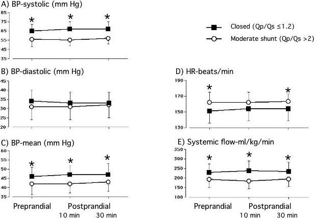
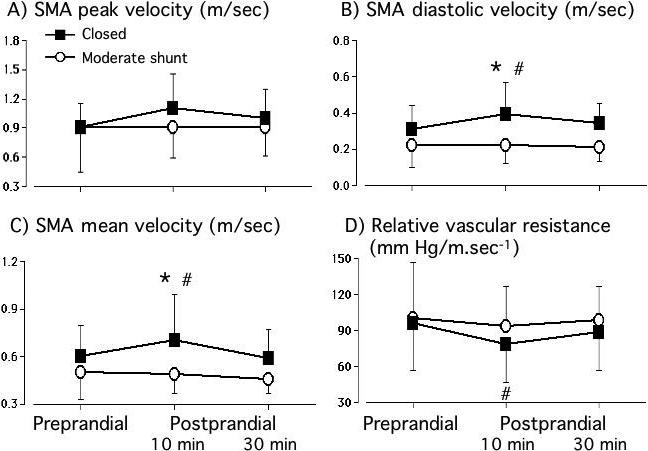
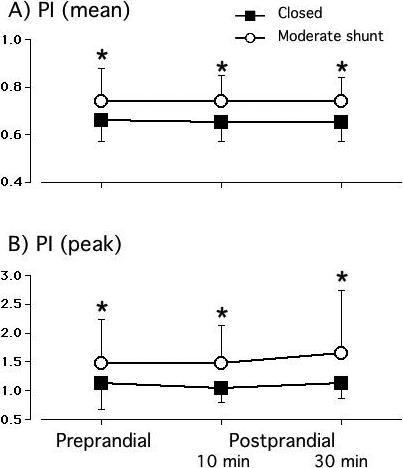
Results: During the preprandial period, baboons with a moderate patent ductus arteriosus had significantly lower blood pressures and systemic blood flows than animals with a closed ductus. Preprandial superior mesenteric artery-blood flow velocities did not differ between the open and closed ductus groups. Animals with a closed ductus increased their superior mesenteric artery-velocities (diastolic and mean) and decreased their superior mesenteric artery relative-vascular-resistance (mean blood pressure/mean superior mesenteric artery-velocity) by 10 minutes after the feeding. By 30 minutes after the feeding, the values were returning to their preprandial values. In contrast, in baboons in the patent ductus arteriosus group, there were no significant changes in superior mesenteric artery-velocity or resistance after the feeding, and superior mesenteric artery-velocities were significantly lower than those in the closed ductus group.
Conclusions: A moderate patent ductus arteriosus shunt limits the ability of the preterm newborn baboon to increase its postprandial mesenteric blood flow velocity. We speculate that this may interfere with its ability to meet increased intestinal metabolic demands and may contribute to feeding difficulties.
Figures
References
-
- Granger HJ, Norris CP. Intrinsic regulation of intestinal oxygenation in the anaesthetized dog. Am J Physiol. 1980;239:H156–H162. - PubMed
-
- Sit SP, Chou CC. Time course of jejunal blood flow, O2 uptake, and O2 extraction during nutrient absorption. Am J Physiol. 1984;247(3 Pt 2):H395–402. - PubMed
-
- Nowicki PT, Miller CE. Autoregulation in the developing postnatal intestinal circulation. Am J Physiol. 1988;254:G189–G193. - PubMed
-
- Crissinger KD, Granger DN. Intestinal blood flow and oxygen consumption: responses to hemorrhage in the developing piglet. Pediatr Res. 1989;26(2):102–5. - PubMed
Publication types
MeSH terms
Grants and funding
LinkOut - more resources
Full Text Sources
Medical
