. 2009 Jan 15;587(1):195-210.
doi: 10.1113/jphysiol.2008.159616. Epub 2008 Nov 10.

An energy supply network of nutrient absorption coordinated by calcium and T1R taste receptors in rat small intestine

Affiliations

An energy supply network of nutrient absorption coordinated by calcium and T1R taste receptors in rat small intestine

Oliver J Mace et al. J Physiol. .

Abstract

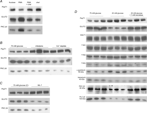
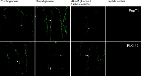
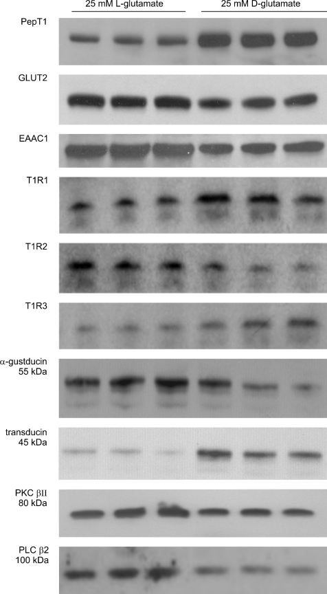
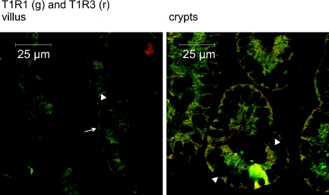
T1R taste receptors are present throughout the gastrointestinal tract. Glucose absorption comprises active absorption via SGLT1 and facilitated absorption via GLUT2 in the apical membrane. Trafficking of apical GLUT2 is rapidly up-regulated by glucose and artificial sweeteners, which act through T1R2 + T1R3/alpha-gustducin to activate PLC beta2 and PKC betaII. We therefore investigated whether non-sugar nutrients are regulated by taste receptors using perfused rat jejunum in vivo. Under different conditions, we observed a Ca(2+)-dependent reciprocal relationship between the H(+)/oligopeptide transporter PepT1 and apical GLUT2, reflecting the fact that trafficking of PepT1 and GLUT2 to the apical membrane is inhibited and activated by PKC betaII, respectively. Addition of L-glutamate or sucralose to a perfusate containing low glucose (20 mM) each activated PKC betaII and decreased apical PepT1 levels and absorption of the hydrolysis-resistant dipeptide L-Phe(PsiS)-L-Ala (1 mM), while increasing apical GLUT2 and glucose absorption within minutes. Switching perfusion from mannitol to glucose (75 mM) exerted similar effects. c-glutamate induced rapid GPCR internalization of T1R1, T1R3 and transducin, whereas sucralose internalized T1R2, T1R3 and alpha-gustducin. We conclude that L-glutamate acts via amino acid and glucose via sweet taste receptors to coordinate regulation of PepT1 and apical GLUT2 reciprocally through a common enterocytic pool of PKC betaII. These data suggest the existence of a wider Ca(2+) and taste receptor-coordinated transport network incorporating other nutrients and/or other stimuli capable of activating PKC betaII and additional transporters, such as the aspartate/glutamate transporter, EAAC1, whose level was doubled by L-glutamate. The network may control energy supply.


Figures

Figure 1
Figure 1. The levels of GLUT2 and PKC βII in the apical membrane show a reciprocal relationship with those of PepT1
Rat jejunum was perfused in KHB for 30 min as described and apical membrane vesicles prepared. Proteins (20 μg) were then separated on 10% SDS-PAGE, transblotted onto PVDF membrane and immunoblotted for GLUT2, PKC βII and PepT1. With the exception of panel A, three separate preparations from two rats each are shown for each condition. All controls, in which antibody has been neutralized by preincubation with excess immunogenic peptide, are described in the references given. The blots for apical GLUT2 and PKC βII are reproduced with permission from the Biochemical Journal (A, Helliwell et al. 2000b), and The Journal of Physiology (B, C and D, Morgan et al. 2007; Mace et al. 2007a,b). The PepT1 blots are new; PepT1 controls were negative (data not shown). A, effect of PMA. Rat jejunum was perfused with 5 mm d-fructose in the presence or absence of 200 nm PMA and/or 2 μm chelerythrin. B, effects of nifedipine and luminal Ca2+ depletion. Jejunum was perfused with 75 mm glucose alone, 75 mm glucose and 10 μm nifedipine or 75 mm glucose in KHB from which Ca2+ was omitted (Ca2+-deplete conditions). C, effect of MLCK inhibition by ML-7. Rat jejunum was perfused with either 75 mm glucose alone or 75 mm glucose and 5 μm ML-7. D, effects glucose and artificial sweetener. Jejunum was perfused with either 75 mm glucose, 20 mm glucose or 20 mm glucose and 1 mm sucralose. Note that the first three samples of the PKC βII were inadvertently placed at the opposite end of the gel; they have been digitally transposed solely for presentational purposes; these PKC βII blots are new.
Figure 2
Figure 2. Immunocytochemistry of PepT1 and PLC β2 regulation by glucose and artificial sweeteners in rat jejunum
Rat jejunum was perfused with 75 mm glucose, 20 mm glucose or 20 mm glucose and 1 mm sucralose for 30 min. Sections (7 μm) were labelled with a rabbit primary antibody detecting either PepT1 or PLC β2 followed by a goat anti-rabbit secondary antibody conjugated to Alexa 488 (green). Arrows: apical membrane; arrowheads: three different types of SCC containing PLC β2. The peptide controls were obtained by neutralization of primary antibody with excess immunogenic peptide. Images were processed using spectral unmixing techniques (see Methods), which automatically subtract all background contributions to leave only specific labelling as demonstrated by the peptide controls, which are totally black.
Figure 3
Figure 3. Transport characteristics of the novel hydrolysis-resistant dipeptide, l-Phe(ΨS)-l-Ala, in rat jejunum
A, rat jejunum was perfused in vivo for 90 min with KHB containing 20 mm glucose and 1 mm l-Phe(ΨS)-l-Ala. The time course depicts the rate of glucose absorption (□, left-hand axis) and rate of l-Phe(ΨS)-l-Ala transport (▪, right hand axis). Rates are measured in μmol min−1 (g dry weight)−1. B, transport characteristics of l-Phe(ΨS)-l-Ala at pH 6.8. The time course of the cumulative appearance of 1 mm l-Phe(ΨS)-l-Ala in the vascular circuit of a luminal and vascularly perfused preparation of rat jejunum in situ. The inset shows the rate of vascular appearance compared with the rat of luminal disappearance. C, specificity of l-Phe(ΨS)-l-Ala for PepT1. Competitive inhibition of the transport of 0.4 μm d-phenylalanine-l-glutamine by l-Phe(ΨS)-l-Ala, when PepT1 was expressed in oocytes.
Figure 4
Figure 4. Regulation of glucose and peptide absorption by intestinal taste receptors
A, regulation by glucose. Jejunum was perfused in vivo with 75 mm mannitol (0 mm glucose) and 1 mm l-Phe(ΨS)-l-Ala for 40 min, when the perfusate was switched (arrow) to an identical one containing 75 mm glucose (0 mm mannitol). The time course depicts the rate of glucose absorption (□, left-hand axis) and the rate of l-Phe(ΨS)-l-Ala transport (▪, right-hand axis). B, regulation by sucralose. Jejunum was perfused in vivo with 20 mm glucose, 1 mm d-Phe(ΨS)-l-Ala and 1 mm sucralose for 40 min, when the perfusate was switched to an otherwise identical perfusate without sucralose. C, regulation by l-glutamate. Jejunum was perfused in vivo with 20 mm glucose, 1 mm l-Phe(ΨS)-l-Ala and 25 mm l-glutamate for 40 min, when the perfusate was changed to an otherwise identical perfusate in which l-glutamate was replaced by d-glutamate.
Figure 5
Figure 5. Regulation of transporters and taste reception signalling components by l-glutamate
Rat jejunum was perfused for 30 min with 20 mm glucose and either 25 mm l-glutamate or 25 mm d-glutamate. Apical membrane vesicles (20 μg) were immunoblotted for the proteins shown.
Figure 6
Figure 6. Colocalization of T1R1 and T1R3 in villus and crypts
Sections (7 μm) were dual-labelled with primary antibodies detecting T1R1 (green) and T1R3 (red) using Alexa 488- and 568-conjugated secondary antibodies. In the merged images for villus, T1R1 and T1R3 are colocalized in the apical membrane (arrow), but not in the basolateral membranes, which appear as dark lines (no labelling, arrowhead) where the membrane separates enterocytes. Note also the contrast with crypts, in which T1R1 and T1R3 are colocalized in the clearly visible basolateral membrane (arrowheads).
Figure 7
Figure 7. An energy supply network of nutrient absorption coordinated by calcium and T1R taste receptors in rat small intestine
The left hand enterocyte depicts the signalling mechanisms for the activation of a common PKC βII pool by Ca2+ absorption and by sweet and amino acid taste receptors; the right hand enterocyte depicts the regulation of transporters by PKC βII.

References

    1. Adler E, Hoon MA, Mueller KL, Chandrashekar J, Ryba NJ, Zuker CS. A novel family of mammalian taste receptors. Cell. 2000;100:693–702. - PubMed
    1. Au A, Gupta A, Schembri P, Cheeseman CI. Rapid insertion of GLUT2 into the rat jejunal brush-border membrane promoted by glucagon-like peptide 2. Biochem J. 2002;367:247–254. - PMC - PubMed
    1. Bailey P. Drug delivery system. 2005. Appl. No. PCT/Gb2005/0001. Pub. No. WO2005/0067978.
    1. Bailey PD, Boyd CA, Collier ID, George JP, Kellett GL, Meredith D, Morgan KM, Pettecrew R, Price RA. Affinity prediction for substrates of the peptide transporter PepT1. Chem Commun (Camb) 2006:323–325. - PubMed
    1. Bailey PD, Boyd CA, Collier ID, Kellett GL, Meredith D, Morgan KM, Pettecrew R, Price RA. Probing dipeptide trans/cis stereochemistry using pH control of thiopeptide analogues, and application to the PepT1 transporter. Org Biomol Chem. 2005;3:4038–4039. - PubMed

Publication types

MeSH terms