. 2009 Jan;77(1):549-56.
doi: 10.1128/IAI.01002-08. Epub 2008 Nov 10.

Characterization of Entamoeba histolytica intermediate subunit lectin-specific human monoclonal antibodies generated in transgenic mice expressing human immunoglobulin loci

Affiliations

Characterization of Entamoeba histolytica intermediate subunit lectin-specific human monoclonal antibodies generated in transgenic mice expressing human immunoglobulin loci

Hiroshi Tachibana et al. Infect Immun. 2009 Jan.

Abstract

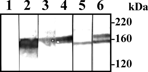
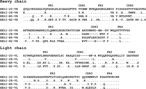
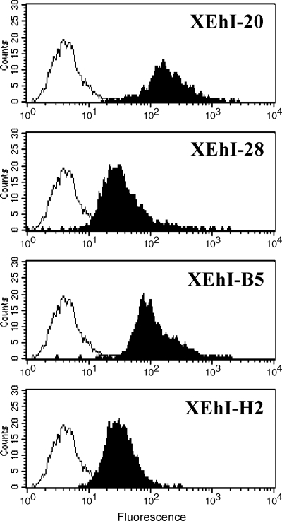
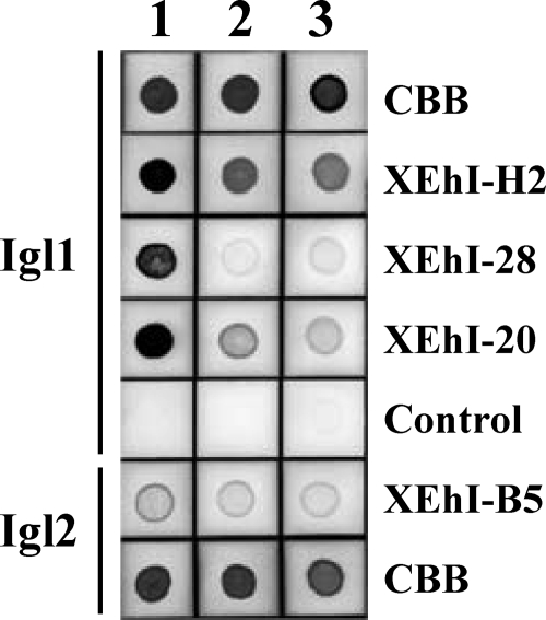
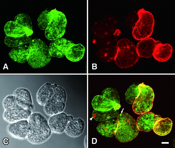
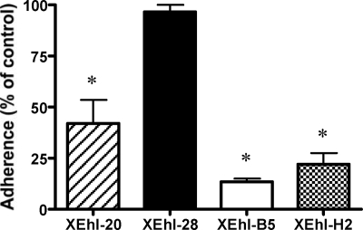
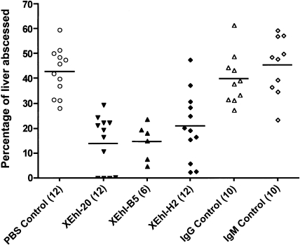
Four fully human monoclonal antibodies (MAbs) to Entamoeba histolytica intermediate subunit lectin (Igl) were prepared in XenoMouse mice, which are transgenic mice expressing human immunoglobulin loci. Examination of the reactivities of these MAbs to recombinant Igl1 and Igl2 of E. histolytica showed that XEhI-20 {immunoglobulin G2(kappa) [IgG2(kappa)]} and XEhI-28 [IgG2(kappa)] were specific to Igl1, XEhI-B5 [IgG2(kappa)] was specific to Igl2, and XEhI-H2 [IgM(kappa)] was reactive with both Igls. Gene analyses revealed that the V(H) and V(L) germ lines were VH3-48 and L2 for XEhI-20, VH3-21 and L2 for XEhI-28, VH3-33 and B3 for XEhI-B5, and VH4-4 and A19 for XEhI-H2, respectively. Flow cytometry analyses showed that the epitopes recognized by all of these MAbs were located on the surfaces of living trophozoites. Confocal microscopy demonstrated that most Igl1 and Igl2 proteins were colocalized on the surface and in the cytoplasm, but different localization patterns in intracellular vacuoles were also present. The preincubation of trophozoites with XEhI-20, XEhI-B5, and XEhI-H2 caused significant inhibition of the adherence of trophozoites to Chinese hamster ovary cells, whereas preincubation with XEhI-28 did not do so. XEhI-20, XEhI-B5, and XEhI-H2 were injected intraperitoneally into hamsters 24 h prior to intrahepatic challenge with E. histolytica trophozoites. One week later, the mean abscess size in groups injected with one of the three MAbs was significantly smaller than that in controls injected with polyclonal IgG or IgM isolated from healthy humans. These results demonstrate that human MAbs to Igls may be applicable for immunoprophylaxis of amebiasis.


Figures

FIG. 1.
FIG. 1.
Western immunoblot analysis of human MAbs to E. histolytica. Lysates of trophozoites from the HM-1:IMSS strain were subjected to SDS-PAGE in a 7.5% gel under nonreducing conditions and transferred onto polyvinylidene difluoride membranes. The strips were treated with the following: lane 1, sera from preimmune XenoMouse mice; lane 2, sera from XenoMouse mice immunized with native E. histolytica Igl; lane 3, XEhI-28; lane 4, XEhI-20; lane 5, XEhI-B5; and lane 6, XEhI-H2. HRP-conjugated goat antibody to human IgG(H+L) was used as a secondary antibody. The numbers on the right indicate molecular masses of size markers.
FIG. 2.
FIG. 2.
Deduced amino acid sequences corresponding to genes coding for heavy- and light-chain variable regions in human MAbs to E. histolytica Igls. FR, framework regions; CDR, complementarity-determining regions. Dashes and dots indicate deletions and identical residues, respectively.
FIG. 3.
FIG. 3.
Flow cytometric analysis of E. histolytica trophozoites stained with human MAbs to Igls (black-filled histograms). Intact trophozoites were incubated with human MAbs XEhI-20, XEhI-28, XEhI-B5, and XEhI-H2, followed by FITC-conjugated goat antibody to human IgG(H+L). The control was stained only with a secondary antibody (unfilled histograms). A representative histogram for each antibody is depicted. Fluorescence levels are expressed in arbitrary units.
FIG. 4.
FIG. 4.
Reactivity of human MAbs to recombinant Igls of E. histolytica in a dot blot analysis. Igl1 and Igl2 (500 ng each) were spotted onto nitrocellulose membranes (lane 1). The same amounts of Igls were spotted after heat treatment under nonreducing (lane 2) and reducing (lane 3) conditions. Two strips were stained with Coomassie brilliant blue (CBB), and other strips were treated with MAbs XEhI-H2, XEhI-28, XEhI-20, and XEhI-B5 or PBS (control). HRP-conjugated goat antibody to human IgG(H+L) was used as a secondary antibody.
FIG. 5.
FIG. 5.
Localization of Igl1 and Igl2 on trophozoites of E. histolytica HM-1:IMSS observed by confocal laser scanning microscopy. Fixed trophozoites were stained with Alexa Fluor 488-labeled XEhI-20, specific for Igl1 (green) (A), and Alexa Fluor 594-labeled XEhI-B5, specific for Igl2 (red) (B). A differential interference contrast microscopy image is shown in panel C. A merged image of panels A and B is shown in panel D. The arrow and arrowhead indicate the individual localization patterns of Igl1 and Igl2, respectively. The bar indicates 10 μm.
FIG. 6.
FIG. 6.
Effects of human MAbs on adherence between E. histolytica and CHO cells. Trophozoites (104) were pretreated with 10 μg of MAb. The rate of adherence is expressed as a percentage of the adherence seen with PBS-treated controls. The results are presented as means ± standard deviations of data from four experiments. Asterisks indicate P values of <0.0001 (for comparison with the PBS control), 0.0029 (for XEhI-20 versus XEhI-B5), 0.0212 (for XEhI-20 versus XEhI-H2), and 0.0284 (for XEhI-B5 versus XEhI-H2).
FIG. 7.
FIG. 7.
Effects of human MAbs on amebic liver abscess formation in hamsters. Each hamster received an intraperitoneal injection of 0.5 ml of PBS containing 5 mg of human MAb 24 h before intrahepatic inoculation with E. histolytica trophozoites. Control hamsters received 0.5 ml of PBS only or 0.5 ml of PBS containing 5 mg of human polyclonal IgG or IgM. Abscess size is expressed as a percentage of the size of the abscessed liver. Horizontal bars indicate the mean values for each group. Numbers in parentheses indicate the number of hamsters in each group. XEhI-20 versus IgG control, P < 0.0001; XEhI-B5 versus IgG control, P < 0.0001; XEhI-H2 versus IgM control, P = 0.0003.

Similar articles

Cited by

References

    1. Abadi, J., J. Friedman, R. A. Mageed, R. Jefferis, M. C. Rodriguez-Barradas, and L. Pirofski. 1998. Human antibodies elicited by a pneumococcal vaccine express idiotypic determinants indicative of V(H)3 gene segment usage. J. Infect. Dis. 178707-716. - PubMed
    1. Andris, J. S., P. H. Ehrlich, L. Ostberg, and J. D. Capra. 1992. Probing the human antibody repertoire to exogenous antigens. Characterization of the H and L chain V region gene segments from anti-hepatitis B virus antibodies. J. Immunol. 1494053-4059. - PubMed
    1. Barbas, C. F., III, E. Bjorling, F. Chiodi, N. Dunlop, D. Cababa, T. M. Jones, S. L. Zebedee, M. A. Persson, P. L. Nara, E. Norrby, et al. 1992. Recombinant human Fab fragments neutralize human type 1 immunodeficiency virus in vitro. Proc. Natl. Acad. Sci. USA 899339-9343. - PMC - PubMed
    1. Bekker, P. J., D. L. Holloway, A. S. Rasmussen, R. Murphy, S. W. Martin, P. T. Leese, G. B. Holmes, C. R. Dunstan, and A. M. DePaoli. 2004. A single-dose placebo-controlled study of AMG 162, a fully human monoclonal antibody to RANKL, in postmenopausal women. J. Bone Miner. Res. 191059-1066. - PubMed
    1. Bird, R. E., K. D. Hardman, J. W. Jacobson, S. Johnson, B. M. Kaufman, S. M. Lee, T. Lee, S. H. Pope, G. S. Riordan, and M. Whitlow. 1988. Single-chain antigen-binding proteins. Science 242423-426. - PubMed

Publication types

MeSH terms

LinkOut - more resources