. 2009 Jan;29(2):389-401.
doi: 10.1128/MCB.00904-08. Epub 2008 Nov 10.

Contributions of F-BAR and SH2 domains of Fes protein tyrosine kinase for coupling to the FcepsilonRI pathway in mast cells

Affiliations

Contributions of F-BAR and SH2 domains of Fes protein tyrosine kinase for coupling to the FcepsilonRI pathway in mast cells

Victor A McPherson et al. Mol Cell Biol. 2009 Jan.

Abstract

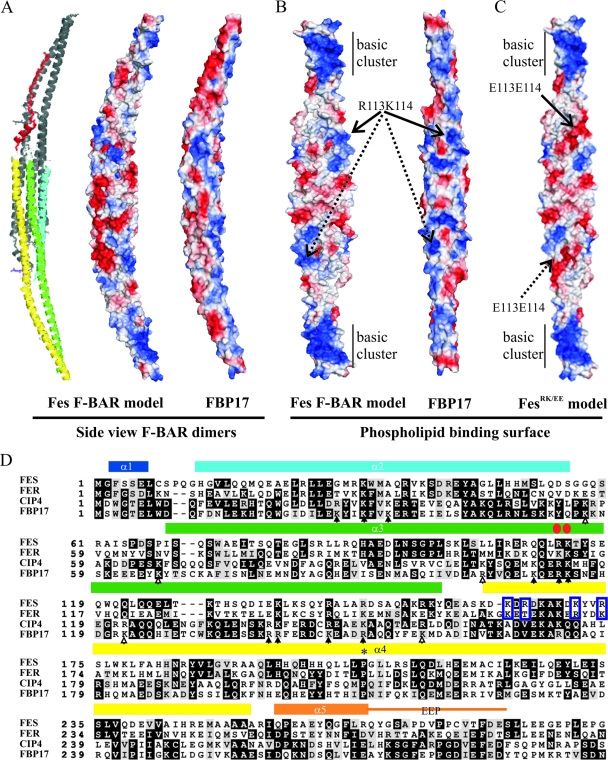
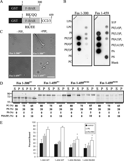
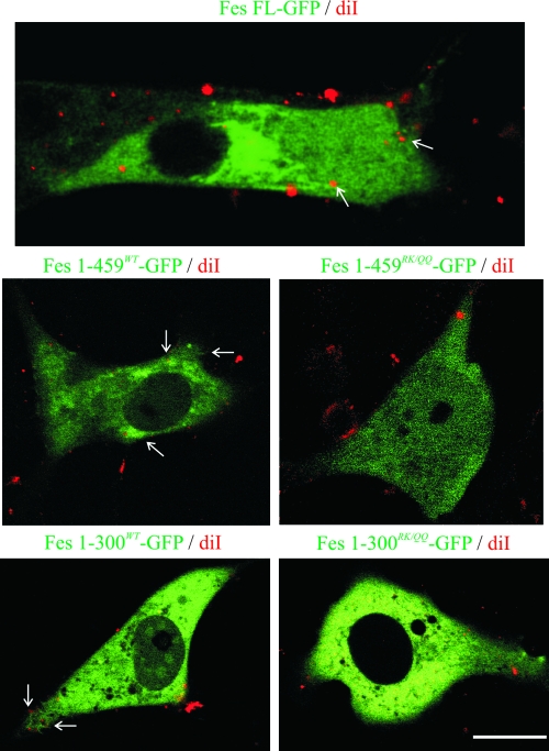
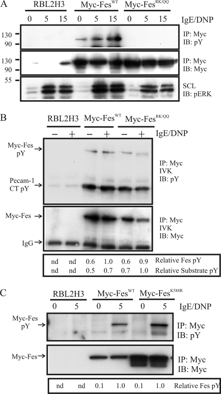
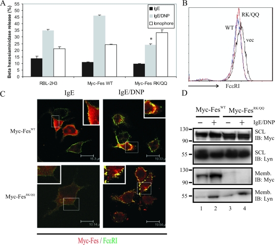
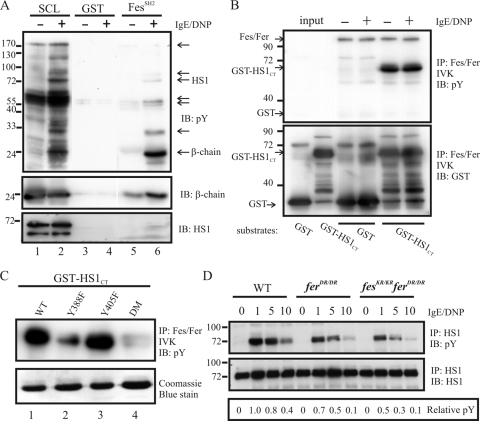
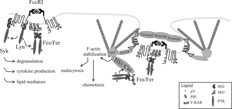
This study investigates the roles of Fer-CIP4 homology (FCH)-Bin/amphiphysin/Rvs (F-BAR) and SH2 domains of Fes protein tyrosine kinase in regulating its activation and signaling downstream of the high-affinity immunoglobulin G (IgE) receptor (FcepsilonRI) in mast cells. Homology modeling of the Fes F-BAR domain revealed conservation of some basic residues implicated in phosphoinositide binding (R113/K114). The Fes F-BAR can bind phosphoinositides and induce tubulation of liposomes in vitro. Mutation of R113/K114 to uncharged residues (RK/QQ) caused a significant reduction in phosphoinositide binding in vitro and a more diffuse cytoplasmic localization in transfected COS-7 cells. RBL-2H3 mast cells expressing full-length Fes carrying the RK/QQ mutation show defects in FcepsilonRI-induced Fes tyrosine phosphorylation and degranulation compared to cells expressing wild-type Fes. This correlated with reduced localization to Lyn kinase-containing membrane fractions for the RK/QQ mutant compared to wild-type Fes in mast cells. The Fes SH2 domain also contributes to Fes signaling in mast cells, via interactions with the phosphorylated FcepsilonRI beta chain and the actin regulatory protein HS1. We show that Fes phosphorylates C-terminal tyrosine residues in HS1 implicated in actin stabilization. Thus, coordinated actions of the F-BAR and SH2 domains of Fes allow for coupling to FcepsilonRI signaling and potential regulation the actin reorganization in mast cells.


Figures

FIG. 1.
FIG. 1.
Homology modeling of the Fes F-BAR domain. The F-BAR domain of human Fes was modeled on the FBP17 F-BAR domain crystal structure (PDB codes 2EFL and 2EFK) (66) as described in Materials and Methods. (A) Fes F-BAR homodimer was generated with the Xtalview program and analyzed in PyMol, and both ribbon and charged-surface representations are shown. Basic residues are shown in blue, and acidic residues are in red for the Fes model and FBP17 structure. (B) The predicted phospholipid binding (concave) faces of the F-BAR domains are shown. The relative positions of conserved residues R113/K114 are indicated by solid and dashed arrows within the Fes F-BAR domain dimer. An more extended patch of basic residues (basic cluster) in Fes compared to FBP17 is indicated near each end of the dimer. (C) A homology model of the FesRK/EE mutant F-BAR domain showing the predicted loss of the central basic residues on the phospholipid-binding face. (D) Multiple sequence alignment of F-BAR domains from human Fes, Fer, CIP4 and FBP17 (from the Baylor College of Medicine Search Launcher using pattern-induced [local] multiple alignment; http://searchlauncher.bcm.tmc.edu/multi-align/Options/pima.html) displayed using the Boxshade server (http://www.ch.embnet.org/software/BOX_form.html). Amino acid numbers are shown on the left, and aligning, identical residues are shaded in black, while conserved residues are shaded gray. The positions of α1 to α5 and the F-BAR extended peptide (EEP) are shown above the residues, based on the structure of FBP17 (59), and are color matched to the ribbon model shown in panel A. Red circles indicate the positions of R113/K114, an asterisk indicates a conserved proline involved in generating F-BAR domain curvature, and blue boxes indicate residues in Fes/Fer PTKs that contribute to the extended basic cluster at the ends of the dimer in panel B. Closed arrows indicate residues in CIP4 shown by Frost et al. to contact lipids of curved membranes, and open arrows indicate residues in CIP4 implicated in binding flat membrane sheets (16).
FIG. 2.
FIG. 2.
Requirement of conserved basic residues for phospholipid binding via the Fes F-BAR domain. (A) Schematic representation of the GST-Fes(1-300) and GST-Fes(1-459) fusion proteins and the positions of RK/QQ and RK/EE mutations. (B) Phospholipid binding screen of the GST-Fes(1-300) and GST-Fes(1-459) proteins with phospholipids arrayed on a filter (PIP Strip; Echelon Bioscience, Inc.). Relative positions of lipid spots are as follows: LPA, lysophosphatidic acid; LPC, lysophosphatidylcholine PI, phosphatidylinositide; PI(3)P, phosphatidylinositol-3-phosphate; PI(4)P, phosphatidylinositol-4-phosphate; PI(5)P, phosphatidylinositol-5-phosphate; S1P, sphingosine-1-phosphate; PI(3,4)P2, phosphatidylinositol-3,4-bisphosphate; PI(3,5)P2, phosphatidylinositol-3,5-bisphosphate; PI(4,5)P2, phosphatidylinositol-4,5-bisphosphate; PI(3,4,5)P3, phosphatidylinositol-3,4,5-trisphosphate; PA, phosphatidic acid. A blank was included. (C) Liposome tubulation assays were carried out with PE/PC/PS-based liposomes, alone or supplemented with PI(4,5)P2, that were incubated with purified Fes(1-300) and Fes(1-459) proteins, as described in Materials and Methods. Representative phase-contrast images are shown. Scale bar, 10 μm. (D) Liposome sedimentation assays using liposomes composed of the indicated combinations of PC, PE, PS, and PI(4,5)P2, incubated with purified GST-Fes(1-300), GST-Fes(1-459)WT, GST-Fes(1-459)RK/QQ, or GST-Fes(1-459)RK/EE protein. Following liposome sedimentation the amount of GST fusion proteins in supernatant (S) and pellet (P) were analyzed by SDS-polyacrylamide gel electrophoresis, followed by Coomassie brilliant blue staining. (E) The results from four to six independent liposome sedimentation assays were quantified by densitometry (graph depicts mean ± standard error of the mean). Asterisks indicate a significant difference (P < 0.05 compared to GST-Fes(1-459)WT by t test).
FIG. 3.
FIG. 3.
The Fes F-BAR domain contributes to tubular localization of Fes in live cells. Live-cell imaging of COS-7 cells transfected with GST-Fes(1-459)WT, GST-Fes(1-459)RK/QQ, and GST-FesFL were incubated with DiIC16(3) (diI) and imaged by confocal microscopy as described in Materials and Methods. Representative confocal microscopy images are shown with overlays of GFP (green) and DiIC16(3)-positive membranes (red). Arrows indicate sites of membrane tubulation. Scale bar, 20 μm. For time-lapse videos showing the dynamic patterns of Fes localization, see the supplemental material.
FIG. 4.
FIG. 4.
The F-BAR domain of Fes is required for FcɛRI-evoked Fes phosphorylation in mast cells. (A) RBL-2H3 cells and derivatives stably expressing FesFL, Myc-FesWT, or Myc-FesRK/QQ were starved and sensitized with anti-DNP-IgE and stimulated for various times (min) with DNP-HSA (100 ng/ml). SCLs were prepared and subjected to either IB with anti-pERK or IP with anti-Myc antibody, followed by IB with anti-pY. The blot was stripped and reprobed with anti-Myc antibody. (B) IVK assays were performed using the same cell lines as above. The relative positions of Myc-Fes autophosphorylation (pY) and substrate phosphorylation (PECAM-1 CT pY) (67) are indicated on the left. The blot was reprobed with anti-Myc to document the relative amounts of Myc-FesWT and Myc-FesRK/QQ in the IVK assay. The position of the IgG heavy chain is indicated on the left. Relative Fes pY and substrate pY were quantified by densitometry. (C) RBL-2H3 cells and derivatives stably expressing Myc-FesWT or the kinase-dead mutant Myc-FesK588R were starved and sensitized with anti-DNP-IgE and stimulated for 5 min with DNP-HSA. Lysates were subjected to IP with anti-Myc and to IB with anti-pY and anti-Myc. The positions of the Myc-Fes proteins are indicated on the left. Relative Fes pY was determined by densitometry. nd, not detected.
FIG. 5.
FIG. 5.
Contributions of Fes F-BAR to regulation of degranulation and localization in RBL-2H3 mast cells. (A) Vector-transfected RBL-2H3 cells and Myc-FesWT- or Myc-FesRK/QQ-expressing cells were sensitized with anti-DNP-IgE and assayed for degranulation in response to antigen or calcium ionophore, as described in Materials and Methods. (B) Surface expression of FcɛRI was determined for RBL-2H3 cells stably transfected with MSCV vector (vec), Myc-FesWT, and Myc-FesRK/QQ. (C) Localization of Myc-FesWT and Myc-FesRK/QQ was analyzed in IgE-sensitized cells (with or without DNP-HSA treatment for 20 min) by immunofluorescence, as described in Materials and Methods. Representative confocal images showing Myc-Fes staining in red and FcɛRI in green are shown (yellow indicates colocalization). Scale bars are shown in the lower right of each panel. (D) Membrane fractions were prepared from Myc-FesWT- and Myc-FesRK/QQ-transfected RBL-2H3 cells as described in Materials and Methods. SCLs and membrane fractions (Memb) were subjected to IB with Myc and Lyn antibodies. Positions of molecular mass markers are indicated on the left.
FIG. 6.
FIG. 6.
Fes SH2 interactions with FcɛRI and its putative substrate HS1 in mast cells. (A) BMMCs were serum starved, sensitized with anti-DNP-IgE, and treated with or without DNP-HSA for 5 min. Lysates were prepared and incubated with purified GST, or GST-FesSH2 fusion protein bound to glutathione-Sepharose beads. Following extensive washing, IB with anti-pY was performed. Relative molecular mass markers are shown on the left, and arrows on the right indicate FesSH2-bound pY-containing proteins. IB performed with anti-β chain and anti-HS1 antibodies on duplicate membranes identified these proteins as potential FesSH2 ligands. (B) IVK assays were performed on Fes/Fer IPs (using a cross-reactive antiserum described previously) (12) from lysates prepared from RBL-2H3 cells sensitized with anti-DNP-IgE and treated with or without DNP-HSA for 2 min. Purified GST and GST-HS1CT were used as substrates in either a mock reaction (input) or Fes/Fer IPs. IVK samples were analyzed by IB with anti-pY and anti-GST antibodies. The positions of autophosphorylated Fes/Fer and of GST proteins are indicated by arrows on the left. Substrates added to each reaction mixture are shown at the bottom. (C) Similar IVK assays were performed for GST-HS1CT fusion proteins harboring the mutations Y388F or Y405F or a double mutant encompassing both mutations (DM). IVK assays were analyzed by IB with anti-pY (upper panel), and substrate amounts are shown by Coomassie brilliant blue staining (lower panel). (D) BMMCs from WT, ferD743R/D743R (ferDR/DR), and fesK588R/K588R ferD743R/D743R (fesKR/KR ferDR/DR) mice were generated, starved of interleukin-3, and sensitized with anti-DNP-IgE. Following treatment with DNP-HSA for the indicated times (min), lysates were prepared and subjected to IP with anti-HS1 and to IB with either anti-pY or anti-HS1. The relative HS1 pY (ratio of pY HS1/total HS1) was determined by densitometry, and values are relative to WT cells treated for 1 min.
FIG. 7.
FIG. 7.
A potential model for contributions of F-BAR-containing adaptor proteins and PTKs in regulating FcɛRI signaling during internalization. FcɛRI-evoked activation of Fes and Fer PTKs involves the actions of an upstream PTK (e.g., Lyn), which is facilitated by F-BAR domain-mediated membrane localization. F-BAR-containing adaptor proteins are known to induce membrane invagination and dynamic actin assembly via SH3-mediated recruitment of WASP and dynamin. We hypothesize that Fes and Fer PTK recruitment of HS1 by FesSH2 and its subsequent phosphorylation contribute stabilization of branched F-actin that promotes endocytosis and chemotaxis of mast cells.

Similar articles

Cited by

References

    1. Abramson, J., and I. Pecht. 2007. Regulation of the mast cell response to the type 1 Fc epsilon receptor. Immunol. Rev. 217231-254. - PubMed
    1. Anderson, D. H., and P. M. Ismail. 1998. v-fps causes transformation by inducing tyrosine phosphorylation and activation of the PDGFβ receptor. Oncogene 162321-2331. - PubMed
    1. Aspenstrom, P., A. Fransson, and N. Richnau. 2006. Pombe Cdc15 homology proteins: regulators of membrane dynamics and the actin cytoskeleton. Trends Biochem. Sci. 31670-679. - PubMed
    1. Balla, T., and P. Varnai. 2002. Visualizing cellular phosphoinositide pools with GFP-fused protein-modules. Sci. STKE 2002PL3. - PubMed
    1. Brooks-Wilson, A. R., E. Ball, and T. Pawson. 1989. The myristylation signal of p60v-src functionally complements the N-terminal fps-specific region of P130gag-fps. Mol. Cell. Biol. 92214-2219. - PMC - PubMed

Publication types

MeSH terms