. 2009 Jan;29(2):602-11.
doi: 10.1128/MCB.01319-08. Epub 2008 Nov 10.

Checkpoint-dependent regulation of origin firing and replication fork movement in response to DNA damage in fission yeast

Affiliations

Checkpoint-dependent regulation of origin firing and replication fork movement in response to DNA damage in fission yeast

Sanjay Kumar et al. Mol Cell Biol. 2009 Jan.

Abstract

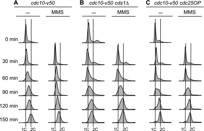
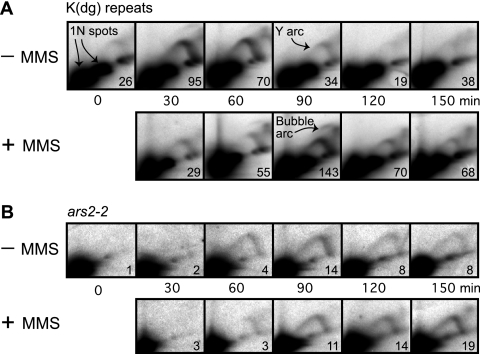
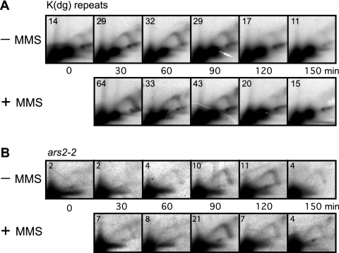
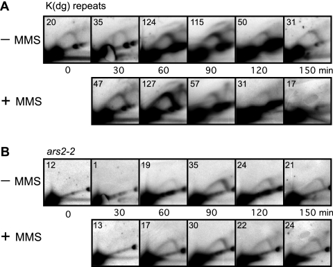
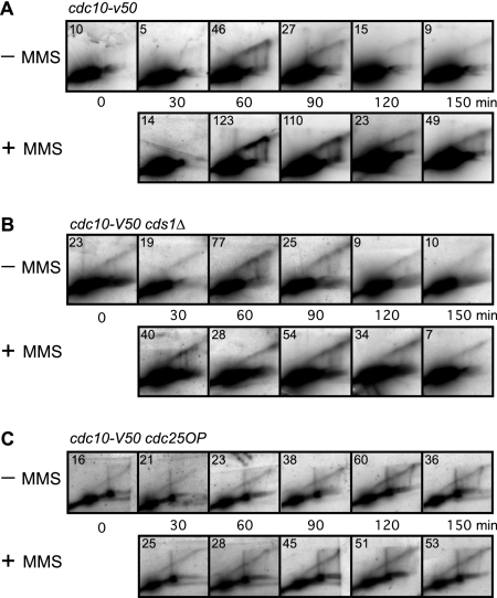
To elucidate the checkpoint mechanism responsible for slowing passage through S phase when fission yeast cells are treated with the DNA-damaging agent methyl methanesulfonate (MMS), we carried out two-dimensional gel analyses of replication intermediates in cells synchronized by cdc10 block (in G(1)) followed by release into synchronous S phase. The results indicated that under these conditions early-firing centromeric origins were partially delayed but late-firing telomeric origins were not delayed. Replication intermediates persisted in MMS-treated cells, suggesting that replication fork movement was inhibited. These effects were dependent on the Cds1 checkpoint kinase and were abolished in cells overexpressing the Cdc25 phosphatase, suggesting a role for the Cdc2 cyclin-dependent kinase. We conclude that both partial inhibition of the firing of a subset of origins and inhibition of replication fork movement contribute to the slowing of S phase in MMS-treated fission yeast cells.


Figures

FIG. 1.
FIG. 1.
Assay for the MMS-induced S-phase damage checkpoint. Cells bearing the cdc10-v50 mutation were arrested in G1 by incubation at 35°C. At 0 min, the cells were released into the cell cycle by reducing the temperature to 25°C. Columns labeled MMS show results for cells treated with 0.015% MMS at 0 min. Samples were collected at the indicated times and analyzed by flow cytometry. (A) Wild-type (cdc10-v50) cells retarded their progression through S phase when treated with MMS. (B and C) In contrast to wild-type cells, cds1Δ (B) and cdc25OP (C) cells failed to slow their progression through S phase upon DNA damage. Because fission yeast cells become elongated when cell cycle arrested, which affects their optical properties, the positions of 1C and 2C peaks cannot be determined from log-phase flow cytometry profiles. We determined the positions of the 1C lines from the position of the major peak at 0 min, modified by the position of the major peak at 30 min in the case (cds1Δ cells) (B) where the 30-min peak was left of the 0-min peak. The positions of the 2C lines were determined by the position of the rightmost of the major peaks (without and with MMS) at 150 min. In all panels, the distances separating the 1C lines from the 2C lines for the untreated and MMS-treated samples are identical. That the untreated cds1Δ cells were indeed at the end of S phase at the 150-min time point, despite showing a peak slightly to the left of the 2C line, is evident from the 2D gels in Fig. 3 and 5B, where the RIs from the untreated cells were consistently at background level at 150 min.
FIG. 2.
FIG. 2.
2D gel analyses of RIs in the wild-type cdc10-v50 strain at the indicated times after shift to 25°C. In each panel, the ratio of RIs to the 1N spot(s) is shown (in units of 10−3) in the lower right corner of each panel. (A) Replication profile of K(dg) repeats. In the absence of MMS these restriction fragments were passively replicated (indicated by strong Y arcs) or, less frequently, were replicated by the firing of internal origins (indicated by faint bubble arcs). The nonreplicating forms of the restriction fragments formed 1N spots. Replication took place predominantly at 30 and 60 min (−MMS row). MMS treatment delayed maximum replication until 90 min, and RIs persisted at 120 and 150 min. (B) Replication profile of ars2-2. The restriction fragment containing ars2-2 replicated later than the K(dg) repeats in the untreated cells, and MMS treatment further delayed its replication. The abundance of RIs in MMS-treated cells increased at the later time points.
FIG. 3.
FIG. 3.
2D gel analyses of RIs in the cds1Δ strain. In each panel, the ratio of RIs to the 1N spot(s) is shown (in units of 10−3) in the upper left corner. MMS treatment failed to delay the replication of either the K(dg) repeats (A) or ars2-2 (B), and RIs did not persist at later time points.
FIG. 4.
FIG. 4.
2D gel analyses of RIs in the cdc25OP strain. In each panel, the ratio of RIs to the 1N spot(s) is shown (in units of 10−3) in the upper left corner. MMS treatment had no significant effect on replication of the K(dg) repeats (A) or ars2-2 (B). The replication profiles appear similar to those for the cds1Δ strain (Fig. 3).
FIG. 5.
FIG. 5.
2D gel analyses of the terminal HindIII fragments at the telomeres of chromosomes 1 and 2 in untreated and MMS-treated cells. In each panel, the ratio of RIs to the 1N spot(s) is shown (in units of 10−3) in the upper left corner. (A) Wild-type cells prolonged their replication of telomeres upon MMS treatment, and the RIs from these regions persisted at later time points. (B and C) Replication of telomeres in cds1Δ (B) and cdc25OP (C) cells was not confined to 60 to 90 min even in the absence of MMS. Upon MMS treatment, the cds1Δ strain (B) showed a further decay of replication synchrony. These telomeric restriction fragments in the cdc25OP strain (C) replicated asynchronously in the absence as well as the presence of MMS.

Similar articles

Cited by

References

    1. Alcasabas, A. A., A. J. Osborn, J. Bacchant, F. Hu, P. J. H. Werler, K. Bousset, K. Furuya, J. F. X. Diffley, A. M. Carr, and S. J. Elledge. 2001. Mrc1 transduces signals of DNA replication stress to activate Rad53. Nat. Cell Biol. 3958-965. - PubMed
    1. Alexandrow, M. G., and J. L. Hamlin. 2005. Chromatin decondensation in S-phase involves recruitment of Cdk2 by Cdc45 and histone H1 phosphorylation. J. Cell Biol. 168875-886. - PMC - PubMed
    1. Andreassen, P. R., G. P. H. Ho, and A. D. D'Andrea. 2006. DNA damage responses and their many interactions with the replication fork. Carcinogenesis 27883-892. - PubMed
    1. Boddy, M. N., and P. Russell. 2001. DNA replication checkpoint. Curr. Biol. 11R953-R956. - PubMed
    1. Brewer, B. J., and W. L. Fangman. 1987. The localization of replication origins on ARS plasmids in S. cerevisiae. Cell 51463-471. - PubMed

Publication types

MeSH terms