. 2009 Jan;29(2):582-601.
doi: 10.1128/MCB.01019-08. Epub 2008 Nov 10.

Nucleus-specific and cell cycle-regulated degradation of mitogen-activated protein kinase scaffold protein Ste5 contributes to the control of signaling competence

Affiliations

Nucleus-specific and cell cycle-regulated degradation of mitogen-activated protein kinase scaffold protein Ste5 contributes to the control of signaling competence

Lindsay S Garrenton et al. Mol Cell Biol. 2009 Jan.

Abstract

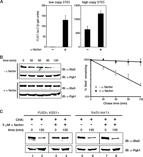
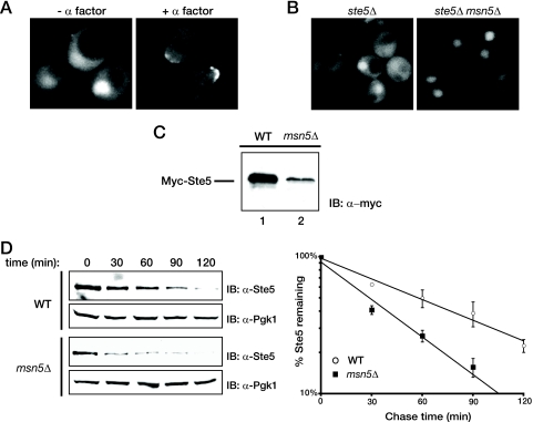
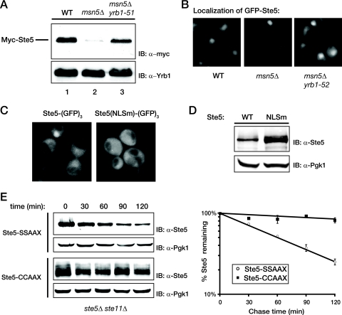
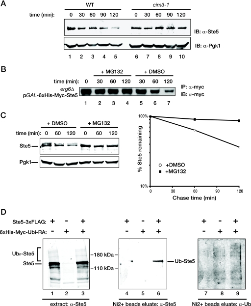
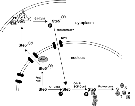
Saccharomyces cerevisiae cells are capable of responding to mating pheromone only prior to their exit from the G(1) phase of the cell cycle. Ste5 scaffold protein is essential for pheromone response because it couples pheromone receptor stimulation to activation of the appropriate mitogen-activated protein kinase (MAPK) cascade. In naïve cells, Ste5 resides primarily in the nucleus. Upon pheromone treatment, Ste5 is rapidly exported from the nucleus and accumulates at the tip of the mating projection via its association with multiple plasma membrane-localized molecules. We found that concomitant with its nuclear export, the rate of Ste5 turnover is markedly reduced. Preventing nuclear export destabilized Ste5, whereas preventing nuclear entry stabilized Ste5, indicating that Ste5 degradation occurs mainly in the nucleus. This degradation is dependent on ubiquitin and the proteasome. We show that Ste5 ubiquitinylation is mediated by the SCF(Cdc4) ubiquitin ligase and requires phosphorylation by the G(1) cyclin-dependent protein kinase (cdk1). The inability to efficiently degrade Ste5 resulted in pathway activation and cell cycle arrest in the absence of pheromone. These findings reveal that maintenance of this MAPK scaffold at an appropriately low level depends on its compartment-specific and cell cycle-dependent degradation. Overall, this mechanism provides a novel means for helping to prevent inadvertent stimulus-independent activation of a response and for restricting and maximizing the signaling competence of the cell to a specific cell cycle stage, which likely works hand in hand with the demonstrated role that G(1) Cdk1-dependent phosphorylation of Ste5 has in preventing its association with the plasma membrane.


Figures

FIG. 1.
FIG. 1.
Effect of Ste5 overexpression on signaling output and of pheromone treatment on Ste5 stability. (A) Exponentially growing cultures of ste5Δ cells (YAB5) carrying a high-copy-number plasmid expressing a FUS1-lacZ reporter gene (YEpU-FUS1Z) and expressing GFP-STE5 from the NOP1 promoter on either a low-copy-number plasmid (left) or a high-copy-number plasmid (right) were treated in the absence (−) or presence (+) of 1 μM α-factor for 60 min, and then the specific activity of β-galactosidase (black columns) was measured {values in Miller units [β-galactosidase (β-gal)] represent the means of three independent trials, and the error bars represent the standard deviations of those means}. The cultures overexpressing GFP-Ste5 grew more slowly and displayed a high proportion of shmoo-shaped cells. (B) (Left) Wild-type (W303) cells were grown to mid-exponential phase and either treated with 3 μM α-factor for 30 min (+ α factor) or not treated with α-factor (− α factor), followed by the addition of CHX (final concentration, 10 μg/ml) to stop any further protein synthesis. Samples were then taken every 30 min as indicated above the gels, and the level of Ste5 was assessed by SDS-PAGE and immunoblotting with rabbit polyclonal anti-Ste5 antiserum (IB: α-Ste5). For a control for equivalent loading in the lanes, the same samples were also immunoblotted with rabbit polyclonal anti-Pgk1 antiserum (IB: α-Pgk1). The results of a representative experiment are shown. (Right) To determine the time dependence of Ste5 degradation, the amount of Ste5 in each sample was quantified using an infrared imaging system (Li-Cor Odyssey version 2.1 software) and normalized to the corresponding content of Pgk1, and the logs of the means of the values so obtained for three independent experiments (performed as described for the left panels) were plotted against time after CHX was added. Error bars represent the standard errors of the means. (C) Wild-type cells (BY4741) or an otherwise isogenic fus3Δ kss1Δ derivative (RCY9321) were grown to mid-exponential phase and either treated (+) or not treated (−) with 3 μM α-factor for 30 min followed by the addition (+) of CHX (10 μg/ml), and 2 h later, a sample was taken and the amounts of Ste5 and Pgk1 were determined as described above for panel B.
FIG. 2.
FIG. 2.
Stabilization of Ste5 is concomitant with its exit from the nucleus. (A) An exponentially growing culture of an ste5Δ mutant (BYB69) expressing Ste5-GFP3 from the STE5 promoter on a CEN plasmid was not treated (− α factor) or treated with 3 μM α-factor (+ α factor). After 45 min, samples of the cells were visualized by standard epifluorescence microscopy as described in Materials and Methods. (B) Exponentially growing cultures of ste5Δ cells (YAB5) or otherwise isogenic ste5Δ msnΔ cells (YAB8 expressing Ste5-GFP3 as in panel A) were visualized by standard fluorescence microscopy. (C) Wild-type (WT) cells (W303) or an otherwise isogenic msn5Δ derivative (HMK30) were transformed with a plasmid expressing Myc-tagged Ste5 from the NOP1 promoter and grown to mid-exponential phase. Extracts were prepared from samples of cell paste (samples with the same weight [wet weight]) by vigorous vortex mixing with glass beads, and the steady-state level of Myc-Ste5 present was analyzed by SDS-PAGE and immunoblotting with an anti-c-Myc monoclonal antibody (IB: α-myc) (MAb 9E10), as described in Materials and Methods. (D) Rate of Ste5 degradation in wild-type cells (W303) or in an otherwise isogenic msn5Δ derivative (HMK30) was monitored after the addition of CHX and plotted as described in the legend to Fig. 1B.
FIG. 3.
FIG. 3.
Retention of Ste5 in the cytosol prevents degradation. (A) Wild-type (WT) cells and otherwise isogenic msn5Δ and msn5Δ yrb1-51 derivatives were transformed with a plasmid expressing Myc-tagged Ste5 from the NOP1 promoter and grown at 23°C until mid-exponential phase, and cell extracts were prepared by vigorous vortex mixing with glass beads. The amount of Ste5 present was assessed by SDS-PAGE and immunoblotting with an anti-c-Myc MAb 9E10 (IB: α-myc). For a control for equivalent loading in the lanes, the same samples were also immunoblotted with rabbit polyclonal anti-Yrb1 antiserum (IB: α-Yrb1). (B) The same cells shown in panel A were transformed with a plasmid expressing GFP-tagged Ste5 from the NOP1 promoter, grown at 23°C until mid-exponential phase, and then visualized by fluorescence microscopy as described in Materials and Methods. Representative fields are shown. (C) Exponentially growing cultures of ste5Δ cells (YAB5) carrying a CEN vector expressing either Ste5-GFP3 or Ste5(NLSm)-GFP3 from the STE5 promoter were visualized by standard fluorescence microscopy. Representative fields are shown. (D) An ste5Δ mutant (BYB69) transformed with a CEN vector expressing either untagged wild-type Ste5 or untagged Ste5(NLSm), each from the STE5 promoter, was grown to mid-exponential phase, harvested, and lysed by the TCA precipitation method as described in Materials and Methods, and the resulting extracts were analyzed by SDS-PAGE and immunoblotting with rabbit polyclonal anti-Ste5 antiserum. For a control for equivalent loading in the lanes, the same samples were also immunoblotted with rabbit polyclonal anti-Pgk1 antiserum. (E) A ste5Δ ste11Δ strain (YLG18) expressing from the GAL1 promoter on a CEN plasmid either Ste5-CCAAX or, as a control, Ste5-SSAAX were pregrown in raffinose-containing medium until early exponential phase, and expression was induced by the addition of galactose. After 2 h, the cells were washed and resuspended in glucose-containing medium to shut off further expression, and samples were taken every 30 min. (Left) The level of Ste5 was determined by immunoblotting with polyclonal anti-Ste5 antiserum, and (right) the results of three independent experiments were plotted as described in the legend to Fig. 1B. For Ste5-CCAAX, the multiple bands represent various states of modification (farnesylation and/or palmitoylation, with or without proteolytic AAX removal accompanied [or not] thereafter by carboxymethylation).
FIG. 4.
FIG. 4.
Ste5 is degraded in a ubiquitin- and proteasome-dependent manner. (A) A wild-type (WT) strain (MHY753) and an otherwise isogenic strain carrying a temperature-sensitive mutation, cim3-1, in the 19S cap of the proteasome (MHY754) were grown to mid-exponential phase at 26°C, shifted to 37°C for 30 min, and treated with CHX, and samples were taken every 30 min, in which the levels of Ste5 and Pgk1 were assessed, as described in the legend to Fig. 1B. (B) BKY48-5C, an erg6Δ mutant permeable to proteasome inhibitor MG132, expressing His6-myc-Ste5 under the control of the GAL1 promoter was grown to mid-exponential phase in raffinose-containing medium, and then Ste5 expression was induced by the addition of galactose. After 2 h, the culture was washed and resuspended in medium containing 2% glucose to shut off any further Ste5 expression (time zero), and half of the culture was incubated with MG132 (final concentration of 50 μM, added from a concentrated stock in DMSO) (+ MG132) or with the same volume of solvent alone (+ DMSO). At the times indicated above the gel, samples were taken, the cells were lysed, and the resulting extracts were subjected to immunoprecipitation using anti-c-Myc MAb 9E10 (IP: α-myc) as described in Materials and Methods. The amount of Myc-tagged Ste5 in the resulting immune complexes was analyzed by SDS-PAGE and immunoblotting with the same antibody (MAb 9E10) (IB: α-myc). (C) To permit entry of MG132 into a wild-type strain (W303), cells pregrown in standard minimal medium supplemented with the appropriate nutrients were incubated for 3 h in a synthetic medium supplemented with 0.1% proline as the nitrogen source and containing 0.003% SDS. One half of the culture received either solvent alone (DMSO) or MG132 (final concentration, 75 μM) in the same solvent as indicated above the gel. After 30 min, CHX (final concentration, 50 μg/ml) was added, and 10 min later, samples were taken at the time points indicated above the gel. The levels of Ste5 and Pgk1 were analyzed (left) and plotted (right) as described in the legend to Fig. 1B. (D) To determine whether Ste5 is a ubiquitinylated protein in vivo during vegetative growth, a ste5Δ mutant (BYB84) was cotransformed with a CEN vector expressing Ste5-FLAG3 from the GAL1 promoter and another CEN vector expressing His6-Myc-Ubi(K48R G76A) from the CUP1 promoter or with each plasmid alone along with the corresponding empty vector as controls. After induction of expression, cells were lysed under denaturing conditions, and a sample of each lysate was analyzed by SDS-PAGE and immunoblotting with polyclonal anti-Ste5 antiserum (lanes 1 to 3). The remaining portion of each lysate was incubated with over Ni2+-loaded NTA beads to enrich for proteins covalently tagged by the His6-Myc-Ubi(K48R G76A). Ubiquitinylated Ste5 (Ub-Ste5) was detected by immunoblotting the eluate of the bead-bound proteins with rabbit polyclonal anti-Ste5 antiserum (lanes 4 to 6), and total ubiquitinylated proteins were detected with a monoclonal antiubiquitin antibody (α-Ub) (lanes 7 to 9).
FIG. 5.
FIG. 5.
RING-H2 domain mutations in Ste5 do not affect its stability. (A) A ste5Δ strain (BYB84) expressing genes under control of the GAL1 promoter on a CEN vector. A strain expressing His6-Myc-tagged versions of either wild-type Ste5 or Ste5(C177A C180A) as indicated above the gels were pregrown in raffinose-containing medium until early exponential phase, and expression was induced by the addition of galactose for 2 h. Cells were then washed and resuspended in glucose-containing medium to shut off further expression, and portions of the culture were taken every 30 min thereafter. These cell samples were lysed by vortex mixing with glass beads and resolved by SDS-PAGE, and the level of Ste5 was determined by immunoblotting with anti-c-myc MAb 9E10 (IB: α-myc). For a control for equivalent loading in the lanes, the same samples were also immunoblotted with rabbit polyclonal anti-Cdc12 antiserum (IB: α-Cdc12). (B) The half-lives of wild-type His6-Myc-tagged versions of wild-type Ste5 or Ste5(C226Y) expressed from the GAL1 promoter on a 2μm DNA plasmid were determined in ste5Δ cells (BYB84) using the GAL shutoff protocol as described above for panel A. (C) The results of the experiment in panel B were quantified, normalized to the level of Cdc12 in the same samples, and plotted on a log scale against time after expression was shut off.
FIG. 6.
FIG. 6.
Ste5 turnover requires the SCFCdc4 ubiquitin ligase. (A) (Left) Exponentially growing cultures of wild-type (WT), msn5Δ, msn5Δ apc10-22, msn5Δ cdc34-2, and msn5Δ cdc4-1 cells were each divided into two equal portions, which were then incubated for 2 h at either a permissive temperature (23°C) or a restrictive temperature (37°C). The cells were then harvested, and protein was extracted by the TCA method. The resulting extracts were resolved by SDS-PAGE and immunoblotted with anti-Ste5 antiserum. (Right) The level of Ste5 was quantified, normalized to the level of Pgk1 in the same samples (not shown), and plotted. Values represent the averages from two independent experiments, and the error bars represent the standard error of those means. (B) (Left) Exponentially growing cultures of wild-type, cdc34-2, or cdc4-1 cells were shifted to 37°C for 45 min followed by the addition of cycloheximide (10 μg/ml) for 10 min. Samples were then taken at the time points indicated above the gels, and the level of endogenous Ste5 was analyzed by SDS-PAGE and immunoblotting with polyclonal anti-Ste5 antiserum. (Right) The level of endogenous Ste5 in wild-type, cdc34-2, or cdc4-1 cells was normalized to the level of Pgk1 in the same samples (not shown) and plotted on a log scale against time after the addition of CHX. Values represent the averages of three independent experiments conducted as shown in the left panels, and the error bars represent the standard errors of those means. (C) Exponentially growing cultures of msn5Δ, msn5Δ cdc34-2, or msn5Δ cdc4-1 cells were shifted to 37°C for 45 min followed by the addition of CHX (10 μg/ml) for 10 min, and then analyzed as explained above for panel B.
FIG. 7.
FIG. 7.
High G1 CDK activity promotes Ste5 degradation. (A) Exponentially growing cultures of an msn5Δ derivative (YLG90) of an wild-type strain (BF264-15D) and an msn5Δ derivative (YLG91) of an otherwise isogenic cdc28-13 strain were divided into two equal portions, which were incubated for 3 h at either the permissive temperature (23°C) or restrictive temperature (37°C). The cells were then harvested, and protein was extracted by the TCA method. The resulting extracts were resolved by SDS-PAGE and immunoblotted with anti-Ste5 antiserum. For a control for equivalent loading in the lanes, the same samples were also immunoblotted with rabbit polyclonal anti-Pgk1 antiserum. (B) Wild-type (WT) cells (BF264-15D) and otherwise isogenic cln1Δ cln2Δ cln3Δ GAL1-CLN2 cells (JTY2142) were pregrown in YP medium with 2% Gal to early exponential phase, at which point each culture was divided into two equal portions. One portion was washed and resuspended in YP medium with 2% Glc to repress Cln2 expression (lanes 2 and 4), and the other portion was maintained in YP medium with 2% Gal (lanes 1 and 3). After 2.5 h, the cells were harvested, and the content of Ste5 and Pgk1 (as a loading control) in each culture was analyzed as described above for panel A. (C) To determine whether the level of Ste5 in the nucleus correlates with cell cycle position, exponentially growing cultures of ste5Δ cells (YAB5) expressing Ste5-GFP3 from the STE5 promoter on a CEN vector were visualized by fluorescence microscopy (∼80% of the cells displayed detectable fluorescence). The pattern of Ste5 localization in representative cells from each phase of the cell cycle, as determined by their budding pattern, is shown. (D) To examine the cell cycle dependence of Ste5 degradation, cells of a cln1Δ cln2Δ cln3Δ GAL1-CLN2 strain or an otherwise isogenic msn5Δ derivative of the same strain were arrested by depletion of the G1 cyclin Cln2 by growth in medium containing 5% Glc for 3 h. Then, the cells were released from G1 arrest by resuspension in medium containing 1% Gal-1% Raf-0.2% Suc. Thereafter, samples were collected at 30 min intervals over the next 2 h, and the content of Ste5 and Pgk1 (as a loading control) in each sample was analyzed as described above for panel A. Measurement of the budding index by examination under the microscope was used to confirm the resumption of cell cycling (n = 200 cells at each time point).
FIG. 8.
FIG. 8.
Ubiquitin-dependent proteolysis of Ste5 prevents inadvertent MAPK signaling. (A) To examine the effect of elevated Ste5 expression when its nuclear degradation is compromised, serial dilutions of exponentially growing wild-type (WT) (W303) and otherwise isogenic cdc34-2, cdc4-1, and apc10-22 mutants carrying either an empty CEN vector or the same vector expressing from the GAL1 promoter either wild-type Ste5 or the signaling-defective mutant Ste5(R407S K411S), were spotted onto plates containing SC medium supplemented with Gal and Ura and grown for 3 days at the indicated temperatures (23 and 30°C). (B) To examine the basis of the growth arrest observed, cultures of cdc34-2 and cdc4-1 cells carrying either an empty vector control (left) of the same plasmid expressing Ste5 from the GAL1 promoter (right) were grown in raffinose-containing at 30°C until early exponential phase, at which time galactose was added (final concentration, 2%). After 5 h, cells were visualized by light microscopy. Each panel depicts a collage of representative cells. (C) The percentages of normal cells, cells with elongated buds, large unbudded (G1-arrested) cells, and overt shmoos in the cell populations shown in panel B were determined by microscopic examination of these cultures (n = 200 cells for each culture).
FIG. 9.
FIG. 9.
Control of Ste5 degradation by compartmentalization and protein kinase action. In naïve cells, Ste5 undergoes nucleocytoplasmic shuttling, with the highest concentration residing in the nucleus. In early G1, prior to the buildup of G1 cyclins (like Cln2) and Cln-dependent activation of Cdk1/Cdc28, the nuclear pool of Ste5 is at a maximum, which can be rapidly drawn upon if the cells encounter mating pheromone. Pheromone-evoked MAPK (Fus3 and Kss1)-dependent phosphorylation of Ste5 stimulates its Msn5-dependent export from the nucleus, and once Ste5 is in the cytosol where it is stable, the amount of Ste5 that can be recruited to the plasma membrane to potentiate signaling becomes correspondingly elevated. This mechanism provides a self-reinforcing feed-forward loop that strengthens and sustains signaling. However, once the levels of the G1 cyclins (especially Cln2) have risen sufficiently, Cdk1(Cdc28) becomes activated. Active Cln-bound Cdk1 promotes the processes required for exit from G1 and entry into S phase (e.g., phosphorylation of the CDK inhibitor Sic1, thereby marking it for its SCFCdc4-dependent and proteasome-mediated destruction). Likewise, active Cln-bound Cdk1 also phosphorylates Ste5; in the cytosol, the G1 Cdk1-dependent modifications block membrane binding, and in the nucleus, G1 Cdk1 phosphorylation initiates SCFCdc4-dependent and proteasome-mediated destruction. Removal of this scaffold protein and its displacement from the membrane obviate the ability of the cells to mount a productive pheromone response at any subsequent stage of the cell division cycle. When the cells return to G1, when Ste5 is stable, an adequate amount of Ste5 builds up and is again available to promote an efficacious response to pheromone, if the cells encounter this signal. See text for further discussion. Abbreviations: PH, pleckstrin homology domain; PM, plasma membrane-binding motif; P, phosphate; NPC, nuclear pore complex: Ub, ubiquitin.

Similar articles

Cited by

References

    1. Alani, E., L. Cao, and N. Kleckner. 1987. A method for gene disruption that allows repeated use of URA3 selection in the construction of multiply disrupted yeast strains. Genetics 116541-545. - PMC - PubMed
    1. Bai, C., P. Sen, K. Hofmann, L. Ma, M. Goebl, J. W. Harper, and S. J. Elledge. 1996. SKP1 connects cell cycle regulators to the ubiquitin proteolysis machinery through a novel motif, the F-box. Cell 86263-274. - PubMed
    1. Bailer, S. M., C. Balduf, J. Katahira, A. Podtelejnikov, C. Rollenhagen, M. Mann, N. Pante, and E. Hurt. 2000. Nup116p associates with the Nup82p-Nsp1p-Nup159p nucleoporin complex. J. Biol. Chem. 27523540-23548. - PubMed
    1. Bardwell, L., J. G. Cook, J. X. Zhu-Shimoni, D. Voora, and J. Thorner. 1998. Differential regulation of transcription: repression by unactivated mitogen-activated protein kinase Kss1 requires the Dig1 and Dig2 proteins. Proc. Natl. Acad. Sci. USA 9515400-15405. - PMC - PubMed
    1. Bashor, C. J., N. C. Helman, S. Yan, and W. A. Lim. 2008. Using engineered scaffold interactions to reshape MAP kinase pathway signaling dynamics. Science 3191539-1543. - PubMed

Publication types

MeSH terms