. 2009 Jan;29(2):378-88.
doi: 10.1128/MCB.01661-08. Epub 2008 Nov 10.

Deletion of Shp2 tyrosine phosphatase in muscle leads to dilated cardiomyopathy, insulin resistance, and premature death

Affiliations

Deletion of Shp2 tyrosine phosphatase in muscle leads to dilated cardiomyopathy, insulin resistance, and premature death

Frederic Princen et al. Mol Cell Biol. 2009 Jan.

Abstract

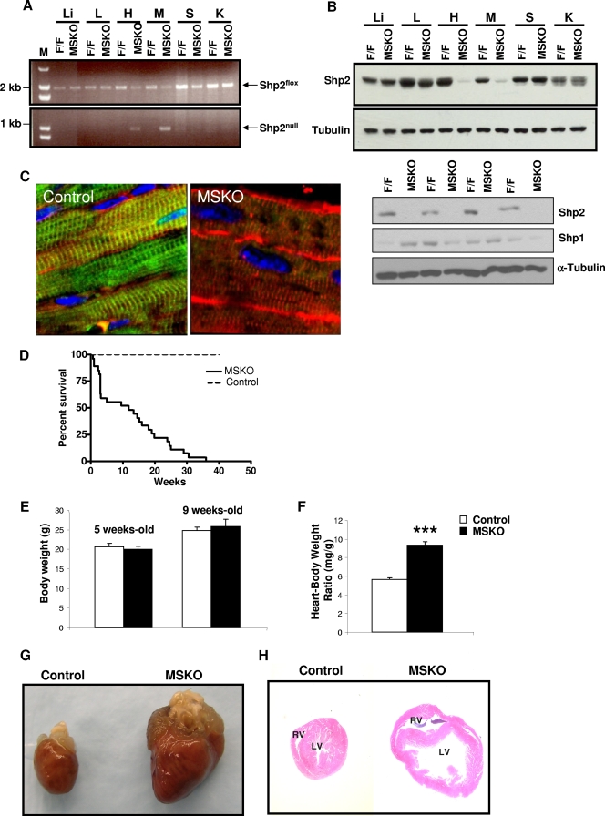
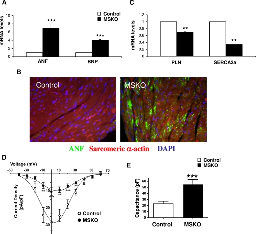
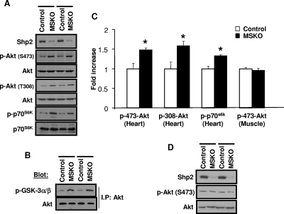
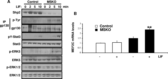
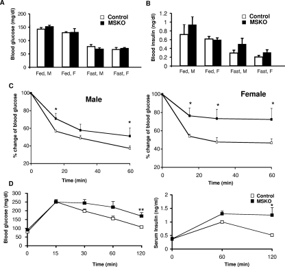
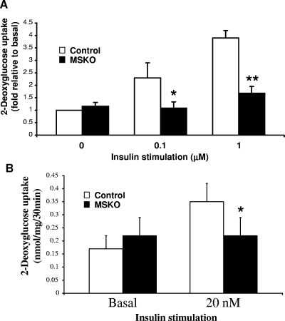
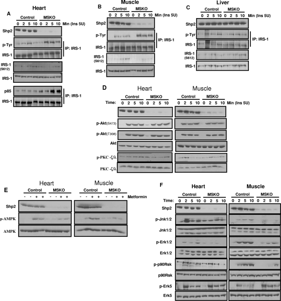
The intracellular signaling mechanisms underlying the pathogenesis of cardiac diseases are not fully understood. We report here that selective deletion of Shp2, an SH2-containing cytoplasmic tyrosine phosphatase, in striated muscle results in severe dilated cardiomyopathy in mice, leading to heart failure and premature mortality. Development of cardiomyopathy in this mouse model is coupled with insulin resistance, glucose intolerance, and impaired glucose uptake in striated muscle cells. Shp2 deficiency leads to upregulation of leukemia inhibitory factor-stimulated phosphatidylinositol 3-kinase/Akt, Erk5, and Stat3 pathways in cardiomyocytes. Insulin resistance and impaired glucose uptake in Shp2-deficient mice are at least in part due to impaired protein kinase C-zeta/lambda and AMP-kinase activities in striated muscle. Thus, we have generated a mouse line modeling human patients suffering from cardiomyopathy and insulin resistance. This study reinforces a concept that a compound disease with multiple cardiovascular and metabolic disturbances can be caused by a defect in a single molecule such as Shp2, which modulates multiple signaling pathways initiated by cytokines and hormones.


Figures

FIG. 1.
FIG. 1.
MSKO mice exhibit dilated cardiomyopathy. (A) PCR detection of Mck-Cre-mediated recombination event in DNA extracts from different tissues. Upper panel, Shp2flox allele; lower panel, Shp2null allele. M, molecular weight marker; Li, liver; L, lung; H, heart; M, skeletal muscle; S, spleen; K, kidney. (B) Immunoblot analysis of Shp2 protein expression in tissues from Shp2flox/flox (control) and MSKO mice. Shp1 expression was examined in skeletal muscles derived from control and MSKO mice with α-tubulin as a loading control (lower panels). (C) Immunofluorescence staining of cardiac left ventricular paraffin sections with antibodies to Shp2 (green) and anti-sarcomeric α-actin (red), as well as counterstained with DAPI (4′,6′-diamidino-2-phenylindole) (blue) (magnification, ×60). (D) Kaplan-Meier survival curves for control and MSKO mice (n = 30). (E) Body weight comparison between 5- and 9-week-old control mice (□) and MSKO mice (▪) (n = 20). (F) Comparison of heart weight/body weight ratios between 5- and 9-week-old control and MSKO mice (n = 13). ***, P < 0.001. (G) Gross morphology of a representative heart from 12-week-old control and MSKO mice. (H) Transversal sections from 12 week-old control and MSKO mouse heart stained with hematoxylin and eosin. The right ventricle (RV) and left ventricle (LV) are shown. All experiments were done on male mice.
FIG. 2.
FIG. 2.
Analysis of cardiac gene expression profile and calcium currents in male MSKO mice. (A) qPCR was performed on total RNA extracted from 4- to 5-week-old control (□) and MSKO (▪) heart tissue. mRNA levels of ANF and BNP normalized against GAPDH are shown (n = 3). (B) Immunofluorescence staining of cardiac left ventricular sections with anti-ANF (green) and anti-sarcomeric α-actin (red) and DAPI (blue) (magnification, ×60). (C) qPCR analysis of mRNA levels of PLN and SERCA2a (n = 3). (D) L-type Ca2+ channel currents (ICa) measurement in postnatal primary cardiomyocyte derived from control (○) and MSKO (•) mice. Voltage-dependent changes in ICa in MSKO (n = 13) and control (n = 14) cells. ICa was normalized to the cell capacitance to yield current densities (pA/pF). (E) Cell capacitance (pF) of control (n = 14) and MSKO (n = 13) cardiomyocytes. *, P < 0.05; **, P < 0.01; ***, P < 0.001.
FIG. 3.
FIG. 3.
Altered Akt activation status in the hearts of MSKO mice. (A) p-Akt (Ser473 and Thr308) and p-p70s6k phosphorylation were measured in 6- to 7-week-old heart lysates. (B) An Akt kinase assay was performed with immunoprecipitated Akt on GSK3 as a substrate, detected using anti-p-GSK3 α/β (Ser21/9). (C) Phosphoprotein levels of p-Akt and p-p70s6k were quantitated relative to the control protein amount (n = 3). *, P < 0.05. (D) p-Akt (Ser473) levels from 6- to 7-week-old skeletal muscle. All experiments were done on male animals.
FIG. 4.
FIG. 4.
Changes in LIF-mediated signaling in cardiomyocytes. (A) LIF (6 × 10⁴ U/kg) was injected into the cava veins of overnight-fasted mice, and heart extracts were immunoblotted with antibodies as indicated. (B) qPCR analysis of MEF2C mRNA levels from 4- to 5-week-old hearts (normalized against GAPDH), with or without LIF stimulation (10 min) (n = 3). **, P < 0.01.
FIG. 5.
FIG. 5.
Metabolic characterization of MSKO mice. (A) Blood glucose measured on randomly fed (fed) or 16-h-fasted (fast) male (M) and female (F) mice (n = 20 to 25). (B) Serum insulin levels were measured in fed and fasted mice (n = 7 to 10). (C) An insulin tolerance test was performed on 6- to 7-week-old male mice and on 6- to 8-week-old female mice (n = 7). (D) A glucose tolerance test was performed on 7- to 8-week-old 16-h-fasted male mice; n = 7. Serum insulin was measured during the glucose tolerance test. *, P < 0.05; **, P < 0.01.
FIG. 6.
FIG. 6.
Glucose uptake in cardiomyocytes and soleus muscle cells. (A) 2-Deoxyglucose uptake in neonatal primary cardiomyocytes. The uptake was determined at baseline and after 30 min of incubation with 0.1 or 1 μM insulin. (B) Soleus muscles were isolated, and glucose uptake was monitored ex vivo at baseline and after 30 min of incubation with 20 nM insulin (n = 3 to 5). *, P < 0.05; **, P < 0.01.
FIG. 7.
FIG. 7.
Insulin-stimulated signaling events. (A to C) Insulin (5 U) was injected into the cava vein, and extracts of heart (A), muscle (B), or liver (C) were subjected to immunoprecipitation or immunoblotting analysis with antibodies as indicated. (D) Tissue samples were collected as in panel A, and immunoblot analyses were done with the indicated antibodies. (E) Metformin (0.4 g/kg) was injected in the cava vein for 5 min. Extracts of heart and muscle were blotted with antibodies as indicated. (F) Insulin (5 U) was injected into the cava veins of overnight fasted mice. Heart and muscle extracts were blotted with antibodies as indicated. The data shown are representative of at least three independent experiments.

Similar articles

Cited by

References

    1. Akazawa, H., and I. Komuro. 2003. Roles of cardiac transcription factors in cardiac hypertrophy. Circ. Res. 921079-1088. - PubMed
    1. Bard-Chapeau, E. A., J. Yuan, N. Droin, S. Long, E. E. Zhang, T. V. Nguyen, and G. S. Feng. 2006. Concerted functions of Gab1 and Shp2 in liver regeneration and hepatoprotection. Mol. Cell. Biol. 264664-4674. - PMC - PubMed
    1. Bruning, J. C., M. D. Michael, J. N. Winnay, T. Hayashi, D. Horsch, D. Accili, L. J. Goodyear, and C. R. Kahn. 1998. A muscle-specific insulin receptor knockout exhibits features of the metabolic syndrome of NIDDM without altering glucose tolerance. Mol. Cell 2559-569. - PubMed
    1. Camper-Kirby, D., S. Welch, A. Walker, I. Shiraishi, K. D. Setchell, E. Schaefer, J. Kajstura, P. Anversa, and M. A. Sussman. 2001. Myocardial Akt activation and gender: increased nuclear activity in females versus males. Circ. Res. 881020-1027. - PubMed
    1. Carroll, R., A. N. Carley, J. R. Dyck, and D. L. Severson. 2005. Metabolic effects of insulin on cardiomyocytes from control and diabetic db/db mouse hearts. Am. J. Physiol. Endocrinol. Metab. 288E900-E906. - PubMed

Publication types

MeSH terms

LinkOut - more resources