. 2009 Jan;15(1):128-37.
doi: 10.1261/rna.1192409. Epub 2008 Nov 10.

Escherichia coli tmRNA lacking pseudoknot 1 tags truncated proteins in vivo and in vitro

Affiliations

Escherichia coli tmRNA lacking pseudoknot 1 tags truncated proteins in vivo and in vitro

Iwona K Wower et al. RNA. 2009 Jan.

Abstract

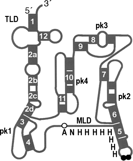
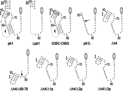
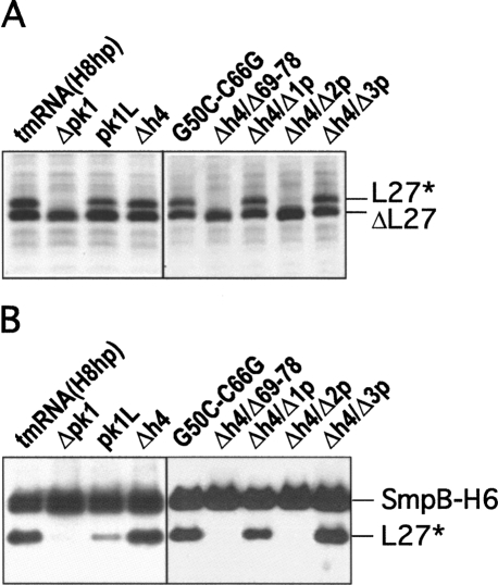
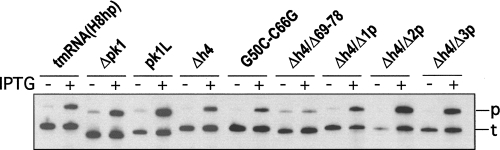
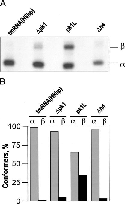
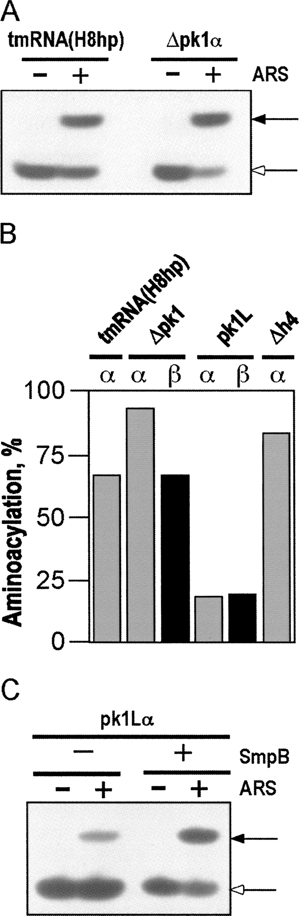
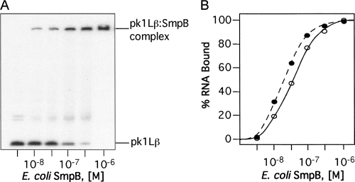
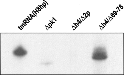
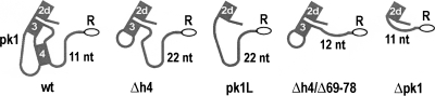
Transfer-messenger RNA (tmRNA) and protein SmpB facilitate trans-translation, a quality-control process that tags truncated proteins with short peptides recognized by a number of proteases and recycles ribosomes stalled at the 3' end of mRNA templates lacking stop codons. The tmRNA molecule is a hybrid of tRNA- and mRNA-like domains that are usually connected by four pseudoknots (pk1-pk4). Replacement of pk1 with a single-stranded RNA yields pk1L, a mutant tmRNA that tags truncated proteins very poorly in vitro but very efficiently in vivo. However, deletion of the whole pk1 is deleterious for protein tagging. In contrast, deletion of helix 4 yields Deltah4, a fully functional tmRNA derivative containing a single hairpin instead of pk1. Further deletions in the pk1 segment yield two subclasses of mutant tmRNAs that are unable to tag truncated proteins, but some of them bind to stalled ribosomes. Our studies demonstrate that pk1 is not essential for tmRNA functions but contributes to the stability of the tmRNA structure. Our studies also indicate that the length of this RNA segment is critical for both tmRNA binding to the ribosome and resumption of translation.


Figures

FIGURE 1.
FIGURE 1.
Structural features of the E. coli tmRNA(H8hp). Schematic representation of the secondary structure of tmRNA(H8hp) composed of the tRNA-like domain (TLD), the mRNA-like domain (MLD) encoding the ANH8 peptide, and four pseudoknots (pk1–pk4). Helices are numbered 1–12. Open and black circles highlight the resume codon (GCA) and two stop codons (UAA–UAA).
FIGURE 2.
FIGURE 2.
Pk1 of the E. coli tmRNA and its mutants. Pk1 is shown with abutting helix 2d. An open square and an open circle denote nucleotides C44 and C66, respectively. Open rectangles highlight mutations of G50C to C50G base pair. Open ovals mark the resume codon (GCA). Arrows indicate deletion sites of nucleotides 54–72 and 69–78 in the pk1L and Δh4/Δ69–78 mutants, respectively. Slanted lines mark deletions of a single base pair, 2 bp, and 3 bp in Δh4/Δ1p, Δh4/Δ2p, and Δh4/Δ3p, respectively. Numbering is according to E. coli tmRNA.
FIGURE 3.
FIGURE 3.
Chemical probing of the E. coli tmRNA(H8hp) derivatives bearing mutations in pk1. Examples of sequencing gels displaying nucleotides modified by DMS in (A) Δpk1, (B) pk1L, and (C) Δh4 mutants. (Lanes C,U,A,G) Sequencing ladders generated by reverse transcription of tmRNA mutants in the presence of ddGTP, ddATP, ddTTP, and ddCTP, respectively. (Lane K) Incubation controls; (lane D) probing under semidenaturing conditions; (lane N) probing under native conditions. Nucleotides modified under native conditions are indicated by black bars both on the autoradiograms and in the secondary structures of the tmRNA mutants.
FIGURE 4.
FIGURE 4.
In vivo and in vitro tagging of truncated ribosomal protein L27 by tmRNA(H8hp) and its derivatives bearing mutations in the pk1. (A) In vivo tagging: Cell lysates from IPTG-induced E. coli IW363 cells coexpressing truncated protein L27, protein SmpB, and mutant tmRNAs were fractionated on a 12.5% SDS-polyacrylamide gel. Tagged proteins were detected by staining the gel with Coomassie Blue. (B) In vitro tagging: Reactions were assembled by addition of circular plasmid pETrpmA-At-1, tmRNA(H8hp) or its mutants, His-tagged E. coli protein SmpB, and E. coli alanyl-tRNA synthetase to the T7 transcription/translation mixture (Promega). After 60 min of incubation at 37°C, His-tagged proteins were captured on Ni-NTA-Sepharose and then fractionated on a 12.5% SDS-polyacrylamide gel. Tagging was visualized by Western blotting with anti-His-tag antibodies. SmpB-H6, ΔL27, and L27* denote His-tagged SmpB, truncated protein L27, and tagged protein L27, respectively.
FIGURE 5.
FIGURE 5.
Processing of tmRNA(H8hp) and its derivatives bearing mutations in the pk1. Northern blot analysis of 1 μg of total RNA extracted from uninduced (−) and IPTG-induced (+) E. coli IW363 cells, fractionated on a 5% denaturing polyacrylamide gel and blotted to a Zeta-probe membrane. [32P]-Labeled oligonucleotide 5′-GGCATCATCATCGT-3′ complementary to E. coli tmRNA residues 125–139 was hybridized to both precursor tmRNAs (p) and mature tmRNAs (t).
FIGURE 6.
FIGURE 6.
Fractionation of tmRNA mutants. (A) TmRNAs were transcribed in vitro, radioactively labeled at the 3′ end by incubation with [α-32P]ATP and ATP/CTP tRNA nucleotidyl transferase, and fractionated by electrophoresis on a native 5% polyacrylamide gel. α and β denote tmRNA conformers. (B) Graphical representation of data derived from the polyacrylamide gel shown in A.
FIGURE 7.
FIGURE 7.
Aminoacylation of tmRNA(H8hp) and its derivatives bearing mutations in the pk1. The aminoacylated and nonaminoacylated [32P]-labeled tmRNAs were digested with RNase T1 to yield CUCCACCA-Ala and CUCCACCA, respectively, and were separated by PAGE according to Varshney et al. (1991). (A) Aminoacylation of tmRNA(H8hp) and Δpk1α (representative examples). (B) Graphical representation of PhosphorImager-derived data from the analysis of aminoacylation of selected tmRNA mutants. (C) Aminoacylation of pk1Lα in the presence and in the absence of His-tagged protein SmpB. Aminoacylation was quantified using a Typhoon PhosphorImager. (+) and (−) indicate the presence and the absence of the alanyl-tRNA synthetase (ARS) and protein SmpB. Solid and open arrows show the positions of [32P]-labeled CUCCACCA-Ala and CUCCACCA, respectively.
FIGURE 8.
FIGURE 8.
Gel-shift analysis of binding the SmpB protein to tmRNAs. The 3′-[32P]-labeled tmRNAs (10−9 M) were titrated with the E. coli SmpB protein. Aliquots of binding mixture were analyzed by electrophoresis on a 5% polyacrylamide gel in TGE buffer. Binding of SmpB to tmRNA derivatives was quantified using a Typhoon PhosphorImager. (A) Gel-shift analysis of pk1Lβ binding to SmpB. (B) Graphical representation of PhosphorImager-derived data illustrating the binding of pkL1β (open circles) and tmRNA(H8hp) (solid circles) to SmpB. Binding curves for other mutant tmRNAs were very similar to the tmRNA(H8hp) curve and therefore are not shown.
FIGURE 9.
FIGURE 9.
Binding of tmRNA(H8hp) and its derivatives bearing mutations in the pk1 to E. coli ribosomes in vivo. Northern blot analysis of 1 μg of RNA extracted from ribosomes isolated from IPTG-induced E. coli IW363 cells expressing either tmRNA(H8hp) or its mutant.
FIGURE 10.
FIGURE 10.
Schematic representation of an RNA segment connecting the TLD and MLD in tmRNAs containing large deletions in the pk1 domain. The length of the connecting RNA segment is expressed as a number of nucleotides. Open ovals and a letter R mark the resume codon (GCA).

References

    1. Andersen E.S., Rosenblad M.A., Larsen N., Westergaard J.C., Burks J., Wower I.K., Wower J., Gorodkin J., Samuelsson T., Zwieb C. The tmRDB and SRPDB resources. Nucleic Acids Res. 2006;34:D163–D168. - PMC - PubMed
    1. Barends S., Wower J., Kraal B. Kinetic parameters for tmRNA binding to alanyl-tRNA synthetase and elongation factor Tu from Escherichia coli . Biochemistry. 2000;39:2652–2658. - PubMed
    1. Barends S., Karzai A.W., Sauer R.T., Wower J., Kraal B. Simultaneous and functional binding of SmpB and EF-Tu-TP to the alanyl acceptor arm of tmRNA. J. Mol. Biol. 2001;314:9–21. - PubMed
    1. Bessho Y., Shibata R., Sekine S., Murayama K., Higashijima K., Hori-Takemoto C., Shirouzu M., Kuramitsu S., Yokoyama S. Structural basis for functional mimicry of long-variable-arm tRNA by transfer-messenger RNA. Proc. Natl. Acad. Sci. 2007;104:8293–8298. - PMC - PubMed
    1. Burks J., Zwieb C., Muller F., Wower I., Wower J. Comparative 3D modeling of tmRNA. BMC Mol. Biol. 2005;6:14–29. - PMC - PubMed

Publication types

LinkOut - more resources