. 2008 Nov 17;183(4):641-51.
doi: 10.1083/jcb.200806065. Epub 2008 Nov 10.

Histone H3 lysine 56 acetylation by Rtt109 is crucial for chromosome positioning

Affiliations

Histone H3 lysine 56 acetylation by Rtt109 is crucial for chromosome positioning

Shin-Ichiro Hiraga et al. J Cell Biol. .

Abstract

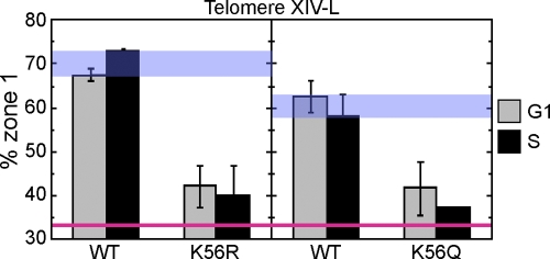
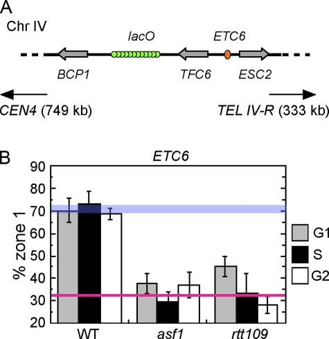
Correct intranuclear organization of chromosomes is crucial for many genome functions, but the mechanisms that position chromatin are not well understood. We used a layered screen to identify Saccharomyces cerevisiae mutants defective in telomere localization to the nuclear periphery. We find that events in S phase are crucial for correct telomere localization. In particular, the histone chaperone Asf1 functions in telomere peripheral positioning. Asf1 stimulates acetylation of histone H3 lysine 56 (H3K56) by the histone acetyltransferase Rtt109. Analysis of rtt109Delta and H3K56 mutants suggests that the acetylation/deacetylation cycle of the H3K56 residue is required for proper telomere localization. The function of H3K56 acetylation in localizing chromosome domains is not confined to telomeres because deletion of RTT109 also prevents the correct peripheral localization of a newly identified S. cerevisiae "chromosome-organizing clamp" locus. Because chromosome positioning is subject to epigenetic inheritance, H3K56 acetylation may mediate correct chromosome localization by facilitating accurate transmission of chromatin status during DNA replication.


Figures

Figure 1.
Figure 1.
Categories of mutants with defective Rap1 organization. The four observed categories of Rap1 organization phenotype are illustrated in the top panels. W, WT-like (i.e., no distinct Rap1 organization defect); A, internal foci; B, diffused fluorescence in entire nucleus; C, larger number of foci mainly still localized at nuclear periphery. Microscopic images on the bottom show strains typical for each category: W, WT; A, ace2Δ; B, ctf18Δ; C, chl1Δ. Bar, 1 μm.
Figure 2.
Figure 2.
Chromosome dot assay identifies a mutant with defective telomere localization to the nuclear periphery. (A) Typical images of WT and rrm3Δ strains with a chromosomal GFP tag 11 kb away from telomere XIV-L, seen as a bright dot. The strains also express Nup49-GFP to visualize nuclear membrane, seen as a dimmer ring. Differential interference contrast (DIC) images are shown on the left. Bars, 2 μm. (B) The zoning of images close to the equatorial plane is used to evaluate telomere localization. Localization of the GFP dot was scored against three concentric zones with equal surface area, as described in Materials and methods. (C) Telomere localization in the identified mutants, assessed separately for cells in G1 phase and S phase. The percentage of cells whose telomere XIV-L dot was peripheral (i.e., in zone 1) is plotted. The red line represents the distribution (33.3%) of a randomly positioned locus. WT values are also indicated (blue bar). For statistical details, see Table II.
Figure 3.
Figure 3.
Telomere delocalization does not cause shortened telomeres, and short telomeres do not preclude peripheral localization. (A) Telomere lengths of telomere localization-defective mutant strains were tested as described in Materials and methods. The result for pgd1Δ is from a different gel performed under identical condition to the others. Positions of molecular weight markers are shown on the left. The black line indicates that lanes have been spliced out. (B) Mean length of the terminal fragment derived from Y′ telomeres plotted against “% zone 1” value of each mutant. For all mutants, the “% zone 1” values differ in G1 and S phases; the lower value is shown. (e.g., for the asf1Δ mutant, “% zone 1” values in G1 and S phase are 67.4% and 43.1%, respectively; the value plotted is 43.1%.)
Figure 4.
Figure 4.
Histone acetyltransferase Rtt109 and histone chaperones Asf1 and Vps75 are crucial for telomere localization. (A) Localization of telomere XIV-L was tested as in Fig. 2. Strains used were GA-1985 (WT), SHY196 (rtt109Δ), SHY173 (asf1Δ), SHY227 (vps75Δ), and SHY229 (asf1Δ vps75Δ). (B) Localization of telomeres VI-R and VIII-L was tested as in Fig. 2. Strains used were GA-1459 (WT) and SHY298 (rtt109Δ) for telomere VI-R, and GA-1986 (WT) and SHY299 (rtt109Δ) for telomere VIII-L. Error bars represent SD of values obtained from independent strain isolates (n = 4 for asf1Δ vps75Δ strain, n = 3 for rtt109Δ strain in A, n = 2 for others). WT and random localization values are indicated by blue and red lines, respectively.
Figure 5.
Figure 5.
Acetylation of H3K56 is crucial for telomere localization. Localization of telomere XIV-L (GFP-tagged 19 kb from telomere) in WT, K56R, and K56Q strains was tested as in Fig. 2. (left) Telomere localization in strains carrying either WT histone H3 allele (SHY248) or K56R allele (SHY247). (right) Telomere localization in strains with either a WT histone H3 allele (SHY310) or K56Q allele (SHY304). Histones H3 and H4 are encoded by only one gene in each strain. Error bars represent SD of values obtained from independent strain isolates (n = 2). Note that the “% zone 1” values shown here for telomere XIV-L are not directly comparable with the other figures because the GFP-tagged locus is different. WT and random localization values are indicated by blue and red lines, respectively. The difference in WT values between left and right panels is presumably caused by the different strain constructs used.
Figure 6.
Figure 6.
Acetylation of H3K56 is crucial for perinuclear localization of ETC6. (A) Strain construct used to test the intranuclear position of ETC6 (located within TFC6-ESC2 intergene on chromosome IV). The neighboring intergene (BCP1-TFC6) was GFP-tagged in WT, asf1Δ, and rtt109Δ mutant strains. The center of the lacO array is 11 kb from the ETC6 locus. (B) Peripheral localization was tested as in Fig. 2. Error bars represent SD of values obtained from independent strain isolates (n = 3, 2, and 4 for the WT, asf1Δ, and rtt109Δ strain, respectively). WT and random localization values are indicated by blue and red lines, respectively.

References

    1. Annunziato, A.T. 2005. Split decision: what happens to nucleosomes during DNA replication? J. Biol. Chem. 280:12065–12068. - PubMed
    1. Annunziato, A.T., and R.L. Seale. 1983. Histone deacetylation is required for the maturation of newly replicated chromatin. J. Biol. Chem. 258:12675–12684. - PubMed
    1. Askree, S.H., T. Yehuda, S. Smolikov, R. Gurevich, J. Hawk, C. Coker, A. Krauskopf, M. Kupiec, and M.J. McEachern. 2004. A genome-wide screen for Saccharomyces cerevisiae deletion mutants that affect telomere length. Proc. Natl. Acad. Sci. USA. 101:8658–8663. - PMC - PubMed
    1. Belgareh, N., and V. Doye. 1997. Dynamics of nuclear pore distribution in nucleoporin mutant yeast cells. J. Cell Biol. 136:747–759. - PMC - PubMed
    1. Brickner, J.H., and P. Walter. 2004. Gene recruitment of the activated INO1 locus to the nuclear membrane. PLoS Biol. 2:e342. - PMC - PubMed

Publication types

MeSH terms