. 2008 Nov 17;183(4):635-40.
doi: 10.1083/jcb.200806112. Epub 2008 Nov 10.

AnkyrinG is required for maintenance of the axon initial segment and neuronal polarity

Affiliations

AnkyrinG is required for maintenance of the axon initial segment and neuronal polarity

Kristian L Hedstrom et al. J Cell Biol. .

Abstract

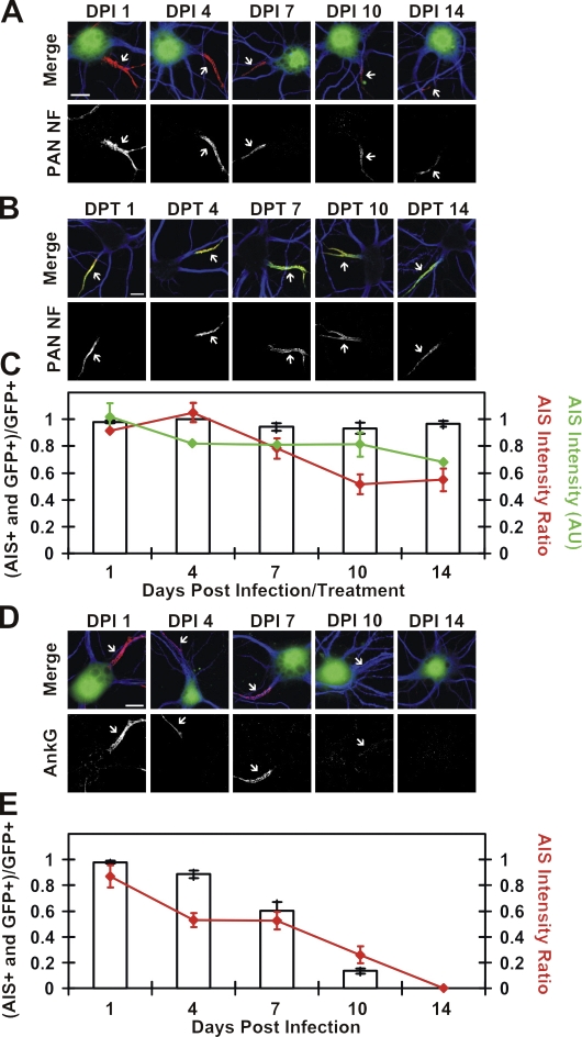
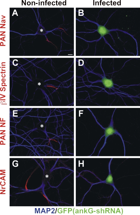
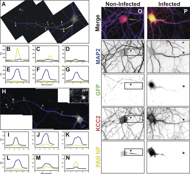
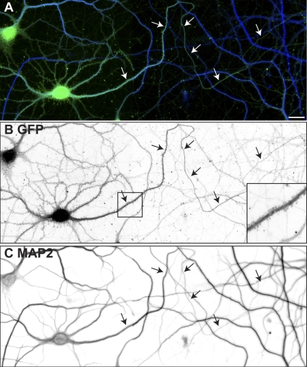
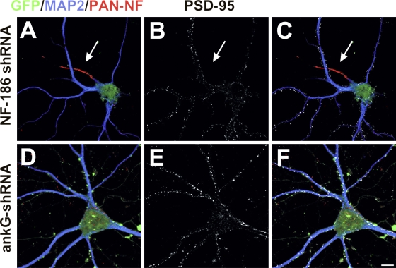
The axon initial segment (AIS) functions as both a physiological and physical bridge between somatodendritic and axonal domains. Given its unique molecular composition, location, and physiology, the AIS is thought to maintain neuronal polarity. To identify the molecular basis of this AIS property, we used adenovirus-mediated RNA interference to silence AIS protein expression in polarized neurons. Some AIS proteins are remarkably stable with half-lives of at least 2 wk. However, silencing the expression of the cytoskeletal scaffold ankyrinG (ankG) dismantles the AIS and causes axons to acquire the molecular characteristics of dendrites. Both cytoplasmic- and membrane-associated proteins, which are normally restricted to somatodendritic domains, redistribute into the former axon. Furthermore, spines and postsynaptic densities of excitatory synapses assemble on former axons. Our results demonstrate that the loss of ankG causes axons to acquire the molecular characteristics of dendrites; thus, ankG is required for the maintenance of neuronal polarity and molecular organization of the AIS.


Figures

Figure 1.
Figure 1.
Stability of NF-186 and ankG at the AIS. (A) NF-186 immunoreactivity (PAN NF, L11A/41.6) in NF-186 shRNA adenovirus–infected (i.e., GFP+) cells at various DPI. (B) Live labeling of neurons using the A12/18.1 mAb (PAN NF) at various DPT. βIV spectrin (green) marks the AIS (merge). (C) Bars indicate the ratio of AIS NF-186–labeled GFP+ neurons to the total number of GFP+ neurons. The mean intensity ratio of a GFP+ cell's AIS to a GFP cell's AIS was calculated per DPI. The red line indicates the mean of the average NF-186 intensity ratio. The green line indicates the mean fluorescence intensity per AIS in A12/18.1 live labeled neurons. AU, arbitrary units. (D) AnkG immunoreactivity in ankG shRNA adenovirus–infected (i.e., GFP+) cells. (E) Bars indicate the ratio of AIS ankG–labeled GFP+ neurons to the total number of GFP+ neurons, and the red line indicates the mean of the average ankG intensity ratio. In A, B, and D, MAP2 (blue) marks somatodendritic domains, and arrows point to the AIS. Error bars represent ±SEM. Bars, 10 μm.
Figure 2.
Figure 2.
AnkG is required for AIS maintenance. Control (A, C, E, and G) and ankG shRNA–infected (B, D, F, and H) neurons immunolabeled for various AIS components: Nav channels (PAN Nav; A and B), βIV spectrin (C and D), NF (PAN NF; E and F), and NrCAM (G and H). GFP fluorescence (green) indicates infected cells, whereas MAP2 (blue) defines somatodendritic domains. Asterisks indicate noninfected neurons. Bar, 10 μm.
Figure 3.
Figure 3.
AnkG is required for maintenance of neuronal polarity. (A) In 20 DIV control neurons, MAP2 (blue) defines somatodendritic domains, whereas PAN NF (yellow) defines the AIS and distal axon. (B–G) Line scans of MAP2 and PAN NF immunofluorescence in A; each line scan shown corresponds to the white lines on the axon (B–D) or dendrite (E–G). (H) In 10 DPI ankG shRNA adenovirus–infected neurons, MAP2 is in all neuronal processes, including the former axon. (I–N) Line scans of MAP2 and PAN NF immunofluorescence shown in H; each line scan shown corresponds to the white lines on the former axon. Distal PAN NF immunofluorescence defines the former axon (N). (O) In 20 DIV control neurons, MAP2 and KCC2 (red) define somatodendritic domains, whereas PAN NF (yellow) defines the axon. Neither MAP2 nor KCC2 is detected in the axon (arrowheads and boxes). (P) In 20 DIV GFP+ ankG shRNA adenovirus–infected neurons, KCC2 and MAP2 immunofluorescence extends into the former axon identified by distal PAN NF immunoreactivity (arrows). AU, arbitrary units. Bars, 10 μm.
Figure 4.
Figure 4.
Loss of ankG causes axons to develop spines. (A) A neuron infected with shRNA to silence the expression of ankG was double labeled for GFP (green) and MAP2 (blue). (B) GFP immunofluorescence shows the axon and the presence of dendritic spines along the axon near the cell body (inset). (C) MAP2 immunoreactivity illustrates high levels of MAP2 along the former axon. In A–C, two adjacent fields were merged to show the distal axon. Arrows point to axons. Bar, 20 μm.
Figure 5.
Figure 5.
Loss of ankG causes axons to acquire excitatory postsynaptic densities. (A–F) Neurons infected with shRNA to silence the expression of NF-186 (A–C) or ankG (D–F) were quadruple labeled for GFP (green), PAN NF (red), MAP2 (blue), and PSD-95 (white). PSD-95 labels excitatory postsynaptic densities and is never found on the axon of NF-186 shRNA adenovirus–infected cells (A–C, arrows). In contrast, all processes of ankG shRNA adenovirus–infected neurons had PSD-95+ puncta, indicating postsynaptic densities on every single process, one of which is a former axon. MAP2 (blue) defines somatodendritic domains, whereas GFP (green) indicates infected neurons. Bar, 10 μm.

References

    1. Arimura, N., and K. Kaibuchi. 2007. Neuronal polarity: from extracellular signals to intracellular mechanisms. Nat. Rev. Neurosci. 8:194–205. - PubMed
    1. Dzhashiashvili, Y., Y. Zhang, J. Galinska, I. Lam, M. Grumet, and J.L. Salzer. 2007. Nodes of Ranvier and axon initial segments are ankyrin G-dependent domains that assemble by distinct mechanisms. J. Cell Biol. 177:857–870. - PMC - PubMed
    1. Garrido, J.J., P. Giraud, E. Carlier, F. Fernandes, A. Moussif, M.P. Fache, D. Debanne, and B. Dargent. 2003. A targeting motif involved in sodium channel clustering at the axonal initial segment. Science. 300:2091–2094. - PubMed
    1. Gomis-Ruth, S., C.J. Wierenga, and F. Bradke. 2008. Plasticity of polarization: changing dendrites into axons in neurons integrated in neuronal circuits. Curr. Biol. 18:992–1000. - PubMed
    1. Grieco, T.M., J.D. Malhotra, C. Chen, L.L. Isom, and I.M. Raman. 2005. Open-channel block by the cytoplasmic tail of sodium channel β4 as a mechanism for resurgent sodium current. Neuron. 45:233–244. - PubMed

Publication types