

Quantitative CT reference values for vertebral trabecular bone density in children and young adults
- PMID: 19001149
- PMCID: PMC2817831
- DOI: 10.1148/radiol.2493080206
Quantitative CT reference values for vertebral trabecular bone density in children and young adults
Abstract
Purpose: To determine normative reference values for vertebral trabecular bone density (TBD) obtained by using quantitative computed tomography (CT) in healthy white children, teenagers, and young adults of both sexes.
Materials and methods: The data presented in this HIPAA-compliant study are a compilation of data from multiple investigations on the determinants of bone acquisition in healthy children conducted at this institution from 1992 to 2006. The institutional review board for clinical investigations approved the protocols for each of these studies, and written informed consent was provided by all parents and/or participants. Quantitative CT measurements of TBD (in milligrams per cubic centimeter) were obtained at the first, second, and third lumbar vertebrae in 1222 healthy white male and female subjects aged 5-21 years (mean age for male subjects, 15.1 years +/- 3.6 [standard deviation]; range, 5.6-21.9 years; mean age for female subjects, 14.2 years +/- 3.9; range, 5.7-21.6 years; mean age for both sexes, 14.6 years +/- 3.8). Mean and standard deviations for TBD were determined for each age group in 1-year intervals, and Student t tests for unpaired data were performed to compare male subjects with female subjects.
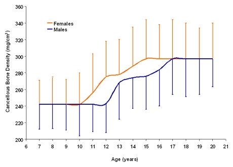
Results: TBD increased equally during growth in male and female subjects. Although the percentage increase in TBD was similar for both sexes (23.7% [57 of 241] for male subjects, 22.2% [54 of 243] for female subjects), the rise began and reached peak values at an earlier age in female subjects; increases in TBD occurred from 10-15 years of age in female subjects, whereas in male subjects, these increases were not observed until age 12 years and were completed at 17 years.
Conclusion: This study provides reference standards for quantitative CT bone measurements in children and young adults, which may aid in the diagnosis, prevention, and treatment of pediatric metabolic bone disorders.
(c) RSNA, 2008.
Figures
References
-
- Mora S, Gilsanz V.Establishment of peak bone mass. Endocrinol Metab Clin North Am 2003;32:39–63 - PubMed
-
- Fares JE, Choucair M, Nabulsi M, Salamoun M, Shahine CH, Fuleihan Gel-H.Effect of gender, puberty, and vitamin D status on biochemical markers of bone remodeling. Bone 2003;33:242–247 - PubMed
-
- Stewart TL, Ralston SH.Role of genetic factors in the pathogenesis of osteoporosis. J Endocrinol 2000;166:235–245 - PubMed
-
- Root AW.Bone strength and the adolescent. Adolesc Med 2002;13:53–72, vi - PubMed
-
- Genant HK, Engelke K, Fuerst T, et al. Noninvasive assessment of bone mineral and structure: state of the art. J Bone Miner Res 1996;11:707–730 - PubMed
Publication types
MeSH terms
Grants and funding
LinkOut - more resources
Full Text Sources
Medical
