

The anatomic correlate of prosopagnosia in semantic dementia
- PMID: 19001253
- PMCID: PMC2676968
- DOI: 10.1212/01.wnl.0000334756.18558.92
The anatomic correlate of prosopagnosia in semantic dementia
Abstract
Objective: To determine the anatomic correlate of prosopagnosia in subjects with semantic dementia.
Methods: We identified all subjects who had been evaluated by an experienced behavioral neurologist, met criteria for semantic dementia, and had completed a volumetric head MRI scan. In all subjects, historical records were reviewed and subjects in which the presence (n = 15) or absence (n = 12) of prosopagnosia was specifically ascertained by the neurologist were identified. Voxel-based morphometry was used to assess patterns of gray matter atrophy in subjects with and without prosopagnosia compared to a group of age and gender-matched normal controls, and compared to each other.
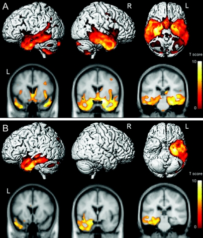
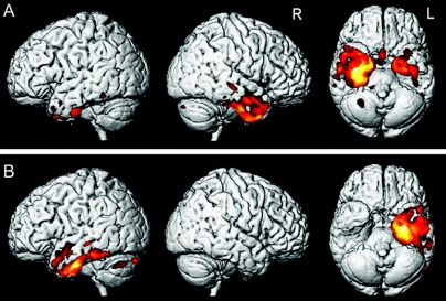
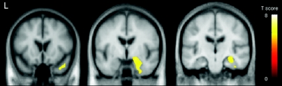
Results: Compared to controls, both groups showed prominent temporal lobe volume loss. Those with prosopagnosia showed bilateral loss but with greater involvement of the right temporal lobe, while those without prosopagnosia showed predominantly left anterior temporal lobe loss. On direct comparison, subjects with prosopagnosia showed greater loss predominantly in the right amygdala, hippocampus, fusiform gyrus, and anterior temporal pole than those without prosopagnosia. No regions were involved to a greater degree in those without prosopagnosia, compared to those with prosopagnosia.
Conclusions: Prosopagnosia appears to be associated with volume loss of the right temporal lobe, particularly medial temporal lobe, fusiform gyrus, and anterior temporal pole, although in semantic dementia it is occurring in the context of bilateral temporal lobe volume loss.
Figures
Similar articles
-
A voxel-based morphometry study of semantic dementia: relationship between temporal lobe atrophy and semantic memory.Ann Neurol. 2000 Jan;47(1):36-45. Ann Neurol. 2000. PMID: 10632099
-
The clinical profile of right temporal lobe atrophy.Brain. 2009 May;132(Pt 5):1287-98. doi: 10.1093/brain/awp037. Epub 2009 Mar 18. Brain. 2009. PMID: 19297506
-
Patterns of temporal lobe atrophy in semantic dementia and Alzheimer's disease.Ann Neurol. 2001 Apr;49(4):433-42. Ann Neurol. 2001. PMID: 11310620
-
Regional deficits in brain volume in schizophrenia: a meta-analysis of voxel-based morphometry studies.Am J Psychiatry. 2005 Dec;162(12):2233-45. doi: 10.1176/appi.ajp.162.12.2233. Am J Psychiatry. 2005. PMID: 16330585 Review.
-
[18F]THK-5351 PET imaging in early-stage semantic variant primary progressive aphasia: a report of two cases and a literature review.BMC Neurol. 2018 Aug 8;18(1):109. doi: 10.1186/s12883-018-1115-3. BMC Neurol. 2018. PMID: 30089453 Free PMC article. Review.
Cited by
-
Gray and white matter water diffusion in the syndromic variants of frontotemporal dementia.Neurology. 2010 Apr 20;74(16):1279-87. doi: 10.1212/WNL.0b013e3181d9edde. Neurology. 2010. PMID: 20404309 Free PMC article.
-
Impairments in the Face-Processing Network in Developmental Prosopagnosia and Semantic Dementia.Cogn Behav Neurol. 2015 Dec;28(4):188-97. doi: 10.1097/WNN.0000000000000077. Cogn Behav Neurol. 2015. PMID: 26705265 Free PMC article.
-
Hemispheric asymmetries in hippocampal volume related to memory in left and right temporal variants of frontotemporal degeneration.Front Neurol. 2024 Apr 29;15:1374827. doi: 10.3389/fneur.2024.1374827. eCollection 2024. Front Neurol. 2024. PMID: 38742046 Free PMC article.
-
A clinical-radiological framework of the right temporal variant of frontotemporal dementia.Brain. 2020 Sep 1;143(9):2831-2843. doi: 10.1093/brain/awaa225. Brain. 2020. PMID: 32830218 Free PMC article.
-
Rapid rate on quasi-speech tasks in the semantic variant of primary progressive aphasia: A non-motor phenomenon?J Acoust Soc Am. 2018 Dec;144(6):3364. doi: 10.1121/1.5082210. J Acoust Soc Am. 2018. PMID: 30599666 Free PMC article.
References
-
- Warrington EK. The selective impairment of semantic memory. Q J Exp Psychol 1975;27:635–657. - PubMed
-
- Snowden JS, Goulding P, Neary D. Semantic dementia: a form of circumscribed cerebral atrophy. Behav Neurol 1989;2:167–182.
-
- Hodges JR, Patterson K, Oxbury S, Funnell E. Semantic dementia. Progressive fluent aphasia with temporal lobe atrophy. Brain 1992;115:1783–1806. - PubMed
-
- Neary D, Snowden JS, Gustafson L, et al. Frontotemporal lobar degeneration: a consensus on clinical diagnostic criteria. Neurology 1998;51:1546–1554. - PubMed
-
- Bozeat S, Lambon Ralph MA, Patterson K, et al. Non-verbal semantic impairment in semantic dementia. Neuropsychologia 2000;38:1207–1215. - PubMed
Publication types
MeSH terms
Grants and funding
LinkOut - more resources
Full Text Sources
Medical
Miscellaneous